


 癌症的基本特征包括细胞增殖、血管生成、迁移、凋亡逃避机制和细胞永生等。找到癌症发生过程中这些通路的关键标记物和对应的抗体用于检测至关重要。
 为您推荐一个泛素化位点预测神器——泛素化分析工具,可以为您的蛋白的泛素化位点作出预测和评分。
 细胞自噬受体图形绘图工具为你的蛋白的细胞受体结合位点作出预测和评分,识别结合到自噬通路中的蛋白是非常重要的,便于让我们理解自噬在正常生理、病理过程中的作用,如发育、细胞分化、神经退化性疾病、压力条件下、感染和癌症。
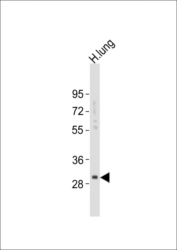
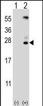
Kallikrein 6 Antibody (N-term)
Purified Rabbit Polyclonal Antibody (Pab)
- 产品详情
- 实验流程
- 背景知识
Application
| WB, IHC-P, FC, E |
|---|---|
| Primary Accession | Q92876 |
| Reactivity | Human |
| Host | Rabbit |
| Clonality | Polyclonal |
| Isotype | Rabbit IgG |
| Calculated MW | 26856 Da |
| Antigen Region | 1-30 aa |
| Gene ID | 5653 |
|---|---|
| Other Names | Kallikrein-6, 3421-, Neurosin, Protease M, SP59, Serine protease 18, Serine protease 9, Zyme, KLK6, PRSS18, PRSS9 |
| Target/Specificity | This Kallikrein 6 antibody is generated from rabbits immunized with a KLH conjugated synthetic peptide between 1-30 amino acids from the N-terminal region of human Kallikrein 6. |
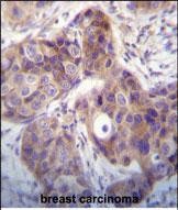
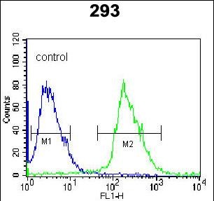
| Dilution | WB~~1:1000 IHC-P~~1:100~500 FC~~1:10~50 E~~Use at an assay dependent concentration. |
| Format | Purified polyclonal antibody supplied in PBS with 0.09% (W/V) sodium azide. This antibody is prepared by Saturated Ammonium Sulfate (SAS) precipitation followed by dialysis against PBS. |
| Storage | Maintain refrigerated at 2-8°C for up to 2 weeks. For long term storage store at -20°C in small aliquots to prevent freeze-thaw cycles. |
| Precautions | Kallikrein 6 Antibody (N-term) is for research use only and not for use in diagnostic or therapeutic procedures. |
| Name | KLK6 |
|---|---|
| Synonyms | PRSS18, PRSS9 |
| Function | Serine protease which exhibits a preference for Arg over Lys in the substrate P1 position and for Ser or Pro in the P2 position. Shows activity against amyloid precursor protein, myelin basic protein, gelatin, casein and extracellular matrix proteins such as fibronectin, laminin, vitronectin and collagen. Degrades alpha-synuclein and prevents its polymerization, indicating that it may be involved in the pathogenesis of Parkinson disease and other synucleinopathies. May be involved in regulation of axon outgrowth following spinal cord injury. Tumor cells treated with a neutralizing KLK6 antibody migrate less than control cells, suggesting a role in invasion and metastasis. |
| Cellular Location | Secreted. Nucleus, nucleolus. Cytoplasm. Mitochondrion. Microsome. Note=In brain, detected in the nucleus of glial cells and in the nucleus and cytoplasm of neurons. Detected in the mitochondrial and microsomal fractions of HEK-293 cells and released into the cytoplasm following cell stress |
| Tissue Location | In fluids, highest levels found in milk of lactating women followed by cerebrospinal fluid, nipple aspirate fluid and breast cyst fluid. Also found in serum, seminal plasma and some amniotic fluids and breast tumor cytosolic extracts. Not detected in urine. At the tissue level, highest concentrations found in glandular tissues such as salivary glands followed by lung, colon, fallopian tube, placenta, breast, pituitary and kidney. Not detected in skin, spleen, bone, thyroid, heart, ureter, liver, muscle, endometrium, testis, pancreas, seminal vesicle, ovary, adrenals and prostate. In brain, detected in gray matter neurons (at protein level). Colocalizes with pathological inclusions such as Lewy bodies and glial cytoplasmic inclusions. Overexpressed in primary breast tumors but not expressed in metastatic tumors. |
For Research Use Only. Not For Use In Diagnostic Procedures.
Provided below are standard protocols that you may find useful for product applications.
BACKGROUND
Kallikreins are a subgroup of serine proteases having diverse physiological functions. Growing evidence suggests that many kallikreins are implicated in carcinogenesis and some have potential as novel cancer and other disease biomarkers. The KLK6 enzyme is regulated by steroid hormones. In tissue culture, the enzyme has been found to generate amyloidogenic fragments from the amyloid precursor protein, suggesting a potential for involvement in Alzheimer's disease.
REFERENCES
Christophi, G.P., et al., J. Neurochem. 91(6):1439-1449 (2004). Bayes, A., et al., Biol. Chem. 385(6):517-524 (2004). Pampalakis, G., et al., Biochem. Biophys. Res. Commun. 320(1):54-61 (2004). Ghosh, M.C., et al., Tumour Biol. 25(4):193-199 (2004). Sauter, E.R., et al., Int. J. Cancer 108(4):588-591 (2004).
终于等到您。ABCEPTA(百远生物)抗体产品。
点击下方“我要评价 ”按钮提交您的反馈信息,您的反馈和评价是我们最宝贵的财富之一,
我们将在1-3个工作日内处理您的反馈信息。
如有疑问,联系:0512-88856768 tech-china@abcepta.com.


