


 癌症的基本特征包括细胞增殖、血管生成、迁移、凋亡逃避机制和细胞永生等。找到癌症发生过程中这些通路的关键标记物和对应的抗体用于检测至关重要。
 为您推荐一个泛素化位点预测神器——泛素化分析工具,可以为您的蛋白的泛素化位点作出预测和评分。
 细胞自噬受体图形绘图工具为你的蛋白的细胞受体结合位点作出预测和评分,识别结合到自噬通路中的蛋白是非常重要的,便于让我们理解自噬在正常生理、病理过程中的作用,如发育、细胞分化、神经退化性疾病、压力条件下、感染和癌症。
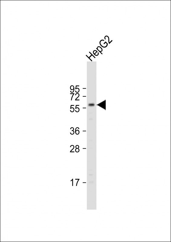
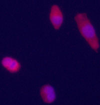
Glypican 3 (GPC3) Antibody (Center)
Purified Rabbit Polyclonal Antibody (Pab)
- 产品详情
- 实验流程
- 背景知识
Application
| IF, WB, E |
|---|---|
| Primary Accession | P51654 |
| Reactivity | Human, Mouse |
| Host | Rabbit |
| Clonality | Polyclonal |
| Isotype | Rabbit IgG |
| Calculated MW | 65563 Da |
| Antigen Region | 335-365 aa |
| Gene ID | 2719 |
|---|---|
| Other Names | Glypican-3, GTR2-2, Intestinal protein OCI-5, MXR7, Secreted glypican-3, GPC3, OCI5 |
| Target/Specificity | This Glypican 3 (GPC3) antibody is generated from rabbits immunized with a KLH conjugated synthetic peptide between 335-365 amino acids from the Central region of human Glypican 3 (GPC3). |
| Dilution | IF~~Tested WB~~1:1000 E~~Use at an assay dependent concentration. |
| Format | Purified polyclonal antibody supplied in PBS with 0.09% (W/V) sodium azide. This antibody is prepared by Saturated Ammonium Sulfate (SAS) precipitation followed by dialysis against PBS. |
| Storage | Maintain refrigerated at 2-8°C for up to 2 weeks. For long term storage store at -20°C in small aliquots to prevent freeze-thaw cycles. |
| Precautions | Glypican 3 (GPC3) Antibody (Center) is for research use only and not for use in diagnostic or therapeutic procedures. |
| Name | GPC3 |
|---|---|
| Synonyms | OCI5 |
| Function | Cell surface proteoglycan (PubMed:14610063). Negatively regulates the hedgehog signaling pathway when attached via the GPI- anchor to the cell surface by competing with the hedgehog receptor PTC1 for binding to hedgehog proteins (By similarity). Binding to the hedgehog protein SHH triggers internalization of the complex by endocytosis and its subsequent lysosomal degradation (By similarity). Positively regulates the canonical Wnt signaling pathway by binding to the Wnt receptor Frizzled and stimulating the binding of the Frizzled receptor to Wnt ligands (PubMed:16227623, PubMed:24496449). Positively regulates the non-canonical Wnt signaling pathway (By similarity). Binds to CD81 which decreases the availability of free CD81 for binding to the transcriptional repressor HHEX, resulting in nuclear translocation of HHEX and transcriptional repression (By similarity). Inhibits the dipeptidyl peptidase activity of DPP4 (PubMed:17549790). Plays a role in limb patterning and skeletal development by controlling the cellular response to BMP4 (By similarity). Modulates the effects of growth factors BMP2, BMP7 and FGF7 on renal branching morphogenesis (By similarity). Required for coronary vascular development (By similarity). Plays a role in regulating cell movements during gastrulation (By similarity). |
| Cellular Location | Cell membrane; Lipid-anchor, GPI-anchor {ECO:0000250|UniProtKB:P13265}; Extracellular side {ECO:0000250|UniProtKB:P13265} |
| Tissue Location | Detected in placenta (at protein level) (PubMed:32337544). Highly expressed in lung, liver and kidney |
For Research Use Only. Not For Use In Diagnostic Procedures.
Provided below are standard protocols that you may find useful for product applications.
BACKGROUND
GPC3 is a cell surface proteoglycan that bears heparan sulfate. This protein may be involved in the suppression/modulation of growth in the predominantly mesodermal tissues and organs, and may play a role in the modulation of IGF2 interactions with its receptor and thereby modulate its function. Members of the glypican-related integral membrane proteoglycan family contain a core protein anchored to the cytoplasmic membrane via a glycosyl phosphatidylinositol (GPI) linkage. These proteins may play a role in the control of cell division, growth regulation, and tumor predisposition. Deletion mutations in GPC3 are the cause of Simpson-Golabi-Behmel syndrome (SGBS), also known as Simpson dysmorphia syndrome (SDYS). SGBS is a condition characterized by pre- and postnatal overgrowth (gigantism) with visceral and skeletal anomalies.
REFERENCES
Nakatsura, T., et al., Clin. Cancer Res. 10(19):6612-6621 (2004).
Boily, G., et al., Br. J. Cancer 90(8):1606-1611 (2004).
Wichert, A., et al., Oncogene 23(4):945-955 (2004).
Midorikawa, Y., et al., Int. J. Cancer 103(4):455-465 (2003).
Sung, Y.K., et al., Cancer Sci. 94(3):259-262 (2003).
终于等到您。ABCEPTA(百远生物)抗体产品。
点击下方“我要评价 ”按钮提交您的反馈信息,您的反馈和评价是我们最宝贵的财富之一,
我们将在1-3个工作日内处理您的反馈信息。
如有疑问,联系:0512-88856768 tech-china@abcepta.com.


