


 癌症的基本特征包括细胞增殖、血管生成、迁移、凋亡逃避机制和细胞永生等。找到癌症发生过程中这些通路的关键标记物和对应的抗体用于检测至关重要。
 为您推荐一个泛素化位点预测神器——泛素化分析工具,可以为您的蛋白的泛素化位点作出预测和评分。
 细胞自噬受体图形绘图工具为你的蛋白的细胞受体结合位点作出预测和评分,识别结合到自噬通路中的蛋白是非常重要的,便于让我们理解自噬在正常生理、病理过程中的作用,如发育、细胞分化、神经退化性疾病、压力条件下、感染和癌症。
Cav3.2 Polyclonal Antibody
- 产品详情
- 实验流程
- 背景知识
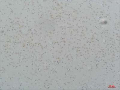
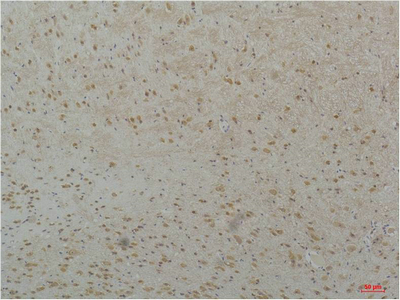
Application
| IHC-P |
|---|---|
| Primary Accession | O95180 |
| Reactivity | Human, Rat, Mouse |
| Host | Rabbit |
| Clonality | Polyclonal |
| Calculated MW | 259163 Da |
| Gene ID | 8912 |
|---|---|
| Other Names | CACNA1H; Voltage-dependent T-type calcium channel subunit alpha-1H; Low-voltage-activated calcium channel alpha1 3.2 subunit; Voltage-gated calcium channel subunit alpha Cav3.2 |
| Dilution | IHC-P~~IHC 1:50-100 |
| Format | Liquid in PBS containing 50% glycerol, 0.5% BSA and 0.09% (W/V) sodium azide. |
| Storage Conditions | -20℃ |
| Name | CACNA1H (HGNC:1395) |
|---|---|
| Function | Voltage-sensitive calcium channel that gives rise to T-type calcium currents. T-type calcium channels belong to the 'low-voltage activated (LVA)' group. A particularity of this type of channel is an opening at quite negative potentials, and a voltage-dependent inactivation (PubMed:27149520, PubMed:9670923, PubMed:9930755). T-type channels serve pacemaking functions in both central neurons and cardiac nodal cells and support calcium signaling in secretory cells and vascular smooth muscle (Probable). They may also be involved in the modulation of firing patterns of neurons (PubMed:15048902). In the adrenal zona glomerulosa, participates in the signaling pathway leading to aldosterone production in response to either AGT/angiotensin II, or hyperkalemia (PubMed:25907736, PubMed:27729216). |
| Cellular Location | Cell membrane; Multi-pass membrane protein. Note=Interaction with STAC increases expression at the cell membrane. |
| Tissue Location | Expressed in the adrenal glomerulosa (at protein level) (PubMed:25907736, PubMed:27729216). In nonneuronal tissues, the highest expression levels are found in the kidney, liver, and heart. In the brain, most abundant in the amygdala, caudate nucleus, and putamen (PubMed:9670923, PubMed:9930755). In the heart, expressed in blood vessels. [Isoform 2]: Expressed in testis, primarily in the germ cells, but not in other portions of the reproductive tract, such as ductus deferens (PubMed:11751928). Not expressed in the brain (PubMed:11751928). |
For Research Use Only. Not For Use In Diagnostic Procedures.
Provided below are standard protocols that you may find useful for product applications.
BACKGROUND
Voltage-sensitive calcium channel that gives rise to T- type calcium currents. T-type calcium channels belong to the "low- voltage activated (LVA)" group. A particularity of this type of channel is an opening at quite negative potentials, and a voltage- dependent inactivation (PubMed:9670923, PubMed:9930755, PubMed:27149520). T-type channels serve pacemaking functions in both central neurons and cardiac nodal cells and support calcium signaling in secretory cells and vascular smooth muscle (Probable). They may also be involved in the modulation of firing patterns of neurons (PubMed:15048902). In the adrenal zona glomerulosa, participates in the signaling pathway leading to aldosterone production in response to either AGT/angiotensin II, or hyperkalemia (PubMed:25907736, PubMed:27729216).
终于等到您。ABCEPTA(百远生物)抗体产品。
点击下方“我要评价 ”按钮提交您的反馈信息,您的反馈和评价是我们最宝贵的财富之一,
我们将在1-3个工作日内处理您的反馈信息。
如有疑问,联系:0512-88856768 tech-china@abcepta.com.


