


 癌症的基本特征包括细胞增殖、血管生成、迁移、凋亡逃避机制和细胞永生等。找到癌症发生过程中这些通路的关键标记物和对应的抗体用于检测至关重要。
 为您推荐一个泛素化位点预测神器——泛素化分析工具,可以为您的蛋白的泛素化位点作出预测和评分。
 细胞自噬受体图形绘图工具为你的蛋白的细胞受体结合位点作出预测和评分,识别结合到自噬通路中的蛋白是非常重要的,便于让我们理解自噬在正常生理、病理过程中的作用,如发育、细胞分化、神经退化性疾病、压力条件下、感染和癌症。
FGF1 Antibody (N-term)
Purified Rabbit Polyclonal Antibody (Pab)
- 产品详情
- 实验流程
- 背景知识
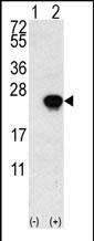
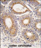
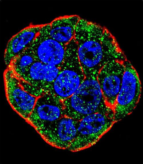
Application
| WB, IHC-P, IF, E |
|---|---|
| Primary Accession | P05230 |
| Other Accession | P20002 |
| Reactivity | Human, Rat, Mouse |
| Predicted | Pig |
| Host | Rabbit |
| Clonality | Polyclonal |
| Isotype | Rabbit IgG |
| Calculated MW | 17460 Da |
| Antigen Region | 5-30 aa |
| Gene ID | 2246 |
|---|---|
| Other Names | Fibroblast growth factor 1, FGF-1, Acidic fibroblast growth factor, aFGF, Endothelial cell growth factor, ECGF, Heparin-binding growth factor 1, HBGF-1, FGF1, FGFA |
| Target/Specificity | This FGF1 antibody is generated from rabbits immunized with a KLH conjugated synthetic peptide between 5-30 amino acids from the N-terminal region of human FGF1. |
| Dilution | WB~~1:1000 IHC-P~~1:100~500 IF~~1:10~50 E~~Use at an assay dependent concentration. |
| Format | Purified polyclonal antibody supplied in PBS with 0.09% (W/V) sodium azide. This antibody is prepared by Saturated Ammonium Sulfate (SAS) precipitation followed by dialysis against PBS. |
| Storage | Maintain refrigerated at 2-8°C for up to 2 weeks. For long term storage store at -20°C in small aliquots to prevent freeze-thaw cycles. |
| Precautions | FGF1 Antibody (N-term) is for research use only and not for use in diagnostic or therapeutic procedures. |
| Name | FGF1 |
|---|---|
| Synonyms | FGFA |
| Function | Plays an important role in the regulation of cell survival, cell division, angiogenesis, cell differentiation and cell migration. Functions as a potent mitogen in vitro. Acts as a ligand for FGFR1 and integrins. Binds to FGFR1 in the presence of heparin leading to FGFR1 dimerization and activation via sequential autophosphorylation on tyrosine residues which act as docking sites for interacting proteins, leading to the activation of several signaling cascades. Binds to integrin ITGAV:ITGB3. Its binding to integrin, subsequent ternary complex formation with integrin and FGFR1, and the recruitment of PTPN11 to the complex are essential for FGF1 signaling. Induces the phosphorylation and activation of FGFR1, FRS2, MAPK3/ERK1, MAPK1/ERK2 and AKT1 (PubMed:18441324, PubMed:20422052). Can induce angiogenesis (PubMed:23469107). |
| Cellular Location | Secreted. Cytoplasm. Cytoplasm, cell cortex. Cytoplasm, cytosol. Nucleus. Note=Lacks a cleavable signal sequence Within the cytoplasm, it is transported to the cell membrane and then secreted by a non-classical pathway that requires Cu(2+) ions and S100A13. Secreted in a complex with SYT1 (By similarity). Binding of exogenous FGF1 to FGFR facilitates endocytosis followed by translocation of FGF1 across endosomal membrane into the cytosol Nuclear import from the cytosol requires the classical nuclear import machinery, involving proteins KPNA1 and KPNB1, as well as LRRC59 |
| Tissue Location | Predominantly expressed in kidney and brain. Detected at much lower levels in heart and skeletal muscle |
For Research Use Only. Not For Use In Diagnostic Procedures.
Provided below are standard protocols that you may find useful for product applications.
BACKGROUND
FGF1 is a member of the fibroblast growth factor (FGF) family. FGF family members possess broad mitogenic and cell survival activities, and are involved in a variety of biological processes, including embryonic development, cell growth, morphogenesis, tissue repair, tumor growth and invasion. This protein functions as a modifier of endothelial cell migration and proliferation, as well as an angiogenic factor. It acts as a mitogen for a variety of mesoderm- and neuroectoderm-derived cells in vitro, thus is thought to be involved in organogenesis.
REFERENCES
Fukushima,S., Int. J. Oncol. 32 (2), 467-473 (2008)
Riley,B.M.Am. J. Med. Genet. A 143 (24), 3228-3234 (2007)
Tomaszewski,M.,Circulation 116 (17), 1915-1924 (2007)
终于等到您。ABCEPTA(百远生物)抗体产品。
点击下方“我要评价 ”按钮提交您的反馈信息,您的反馈和评价是我们最宝贵的财富之一,
我们将在1-3个工作日内处理您的反馈信息。
如有疑问,联系:0512-88856768 tech-china@abcepta.com.


