


 癌症的基本特征包括细胞增殖、血管生成、迁移、凋亡逃避机制和细胞永生等。找到癌症发生过程中这些通路的关键标记物和对应的抗体用于检测至关重要。
 为您推荐一个泛素化位点预测神器——泛素化分析工具,可以为您的蛋白的泛素化位点作出预测和评分。
 细胞自噬受体图形绘图工具为你的蛋白的细胞受体结合位点作出预测和评分,识别结合到自噬通路中的蛋白是非常重要的,便于让我们理解自噬在正常生理、病理过程中的作用,如发育、细胞分化、神经退化性疾病、压力条件下、感染和癌症。
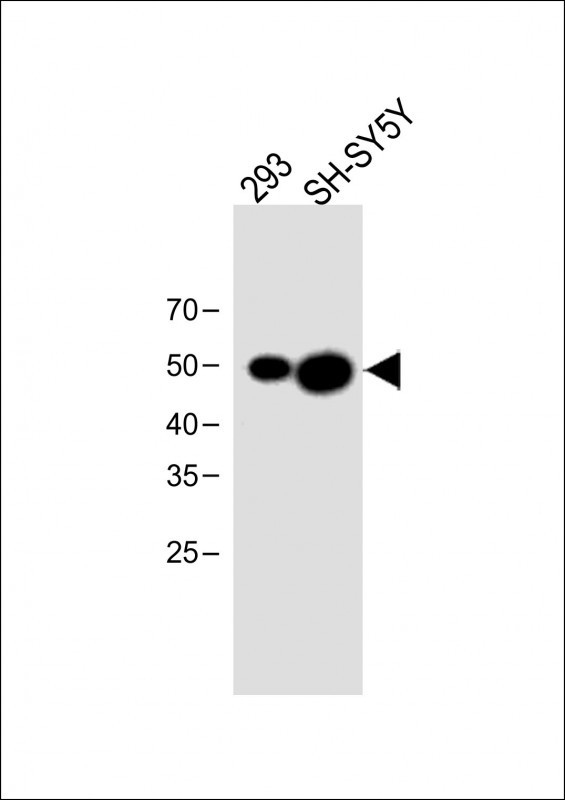
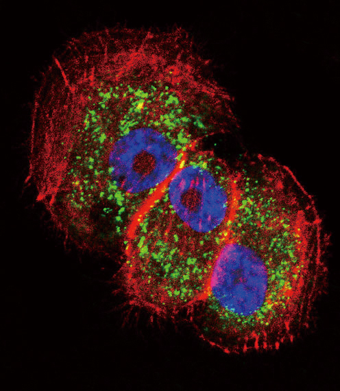
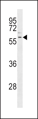
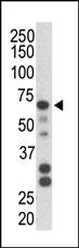
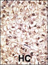
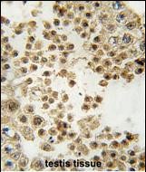
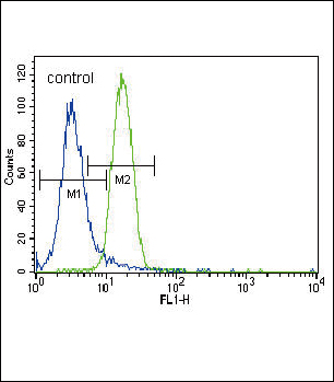
Parkin Antibody (C-term)
Purified Rabbit Polyclonal Antibody (Pab)
- 产品详情
- 实验流程
- 背景知识
Application
| WB, IHC-P, IF, FC, E |
|---|---|
| Primary Accession | O60260 |
| Other Accession | NP_004553 |
| Reactivity | Human, Mouse |
| Host | Rabbit |
| Clonality | Polyclonal |
| Isotype | Rabbit IgG |
| Calculated MW | 51641 Da |
| Antigen Region | 387-417 aa |
| Gene ID | 5071 |
|---|---|
| Other Names | E3 ubiquitin-protein ligase parkin, 632-, Parkinson juvenile disease protein 2, Parkinson disease protein 2, PARK2, PRKN |
| Target/Specificity | This Parkin antibody is generated from rabbits immunized with a KLH conjugated synthetic peptide between 387-417 amino acids from the C-terminal region of human Parkin. |
| Dilution | WB~~1:1000 IHC-P~~1:100~500 IF~~1:10~50 FC~~1:10~50 E~~Use at an assay dependent concentration. |
| Format | Purified polyclonal antibody supplied in PBS with 0.05% (V/V) Proclin 300. This antibody is purified through a protein A column, followed by peptide affinity purification. |
| Storage | Maintain refrigerated at 2-8°C for up to 2 weeks. For long term storage store at -20°C in small aliquots to prevent freeze-thaw cycles. |
| Precautions | Parkin Antibody (C-term) is for research use only and not for use in diagnostic or therapeutic procedures. |
| Name | PRKN (HGNC:8607) |
|---|---|
| Synonyms | PARK2 |
| Function | Functions within a multiprotein E3 ubiquitin ligase complex, catalyzing the covalent attachment of ubiquitin moieties onto substrate proteins (PubMed:10888878, PubMed:10973942, PubMed:11431533, PubMed:12150907, PubMed:12628165, PubMed:15105460, PubMed:16135753, PubMed:21376232, PubMed:21532592, PubMed:22396657, PubMed:23620051, PubMed:23754282, PubMed:24660806, PubMed:24751536, PubMed:29311685, PubMed:32047033). Substrates include SYT11 and VDAC1 (PubMed:29311685, PubMed:32047033). Other substrates are BCL2, CCNE1, GPR37, RHOT1/MIRO1, MFN1, MFN2, STUB1, SNCAIP, SEPTIN5, TOMM20, USP30, ZNF746, MIRO1 and AIMP2 (PubMed:10888878, PubMed:10973942, PubMed:11431533, PubMed:12150907, PubMed:12628165, PubMed:15105460, PubMed:16135753, PubMed:21376232, PubMed:21532592, PubMed:22396657, PubMed:23620051, PubMed:23754282, PubMed:24660806, PubMed:24751536). Mediates monoubiquitination as well as 'Lys-6', 'Lys-11', 'Lys-48'-linked and 'Lys-63'-linked polyubiquitination of substrates depending on the context (PubMed:19229105, PubMed:20889974, PubMed:25474007, PubMed:25621951, PubMed:32047033). Participates in the removal and/or detoxification of abnormally folded or damaged protein by mediating 'Lys-63'-linked polyubiquitination of misfolded proteins such as PARK7: 'Lys-63'-linked polyubiquitinated misfolded proteins are then recognized by HDAC6, leading to their recruitment to aggresomes, followed by degradation (PubMed:17846173, PubMed:19229105). Mediates 'Lys-63'-linked polyubiquitination of a 22 kDa O-linked glycosylated isoform of SNCAIP, possibly playing a role in Lewy-body formation (PubMed:11431533, PubMed:11590439, PubMed:15105460, PubMed:15728840, PubMed:19229105). Mediates monoubiquitination of BCL2, thereby acting as a positive regulator of autophagy (PubMed:20889974). Protects against mitochondrial dysfunction during cellular stress, by acting downstream of PINK1 to coordinate mitochondrial quality control mechanisms that remove and replace dysfunctional mitochondrial components (PubMed:11439185, PubMed:18957282, PubMed:19029340, PubMed:19966284, PubMed:21376232, PubMed:22082830, PubMed:22396657, PubMed:23620051, PubMed:23933751, PubMed:24660806, PubMed:24784582, PubMed:24896179, PubMed:25474007, PubMed:25527291, PubMed:32047033). Depending on the severity of mitochondrial damage and/or dysfunction, activity ranges from preventing apoptosis and stimulating mitochondrial biogenesis to regulating mitochondrial dynamics and eliminating severely damaged mitochondria via mitophagy (PubMed:11439185, PubMed:19029340, PubMed:19801972, PubMed:19966284, PubMed:21376232, PubMed:22082830, PubMed:22396657, PubMed:23620051, PubMed:23685073, PubMed:23933751, PubMed:24896179, PubMed:25527291, PubMed:32047033, PubMed:33499712). Activation and recruitment onto the outer membrane of damaged/dysfunctional mitochondria (OMM) requires PINK1-mediated phosphorylation of both PRKN and ubiquitin (PubMed:24660806, PubMed:24784582, PubMed:25474007, PubMed:25527291). After mitochondrial damage, functions with PINK1 to mediate the decision between mitophagy or preventing apoptosis by inducing either the poly- or monoubiquitination of VDAC1, respectively; polyubiquitination of VDAC1 promotes mitophagy, while monoubiquitination of VDAC1 decreases mitochondrial calcium influx which ultimately inhibits apoptosis (PubMed:27534820, PubMed:32047033). When cellular stress results in irreversible mitochondrial damage, promotes the autophagic degradation of dysfunctional depolarized mitochondria (mitophagy) by promoting the ubiquitination of mitochondrial proteins such as TOMM20, RHOT1/MIRO1, MFN1 and USP30 (PubMed:19029340, PubMed:19966284, PubMed:21753002, PubMed:22396657, PubMed:23620051, PubMed:23685073, PubMed:23933751, PubMed:24896179, PubMed:25527291). Preferentially assembles 'Lys-6'-, 'Lys-11'- and 'Lys-63'-linked polyubiquitin chains, leading to mitophagy (PubMed:25621951, PubMed:32047033). The PINK1-PRKN pathway also promotes fission of damaged mitochondria by PINK1-mediated phosphorylation which promotes the PRKN-dependent degradation of mitochondrial proteins involved in fission such as MFN2 (PubMed:23620051). This prevents the refusion of unhealthy mitochondria with the mitochondrial network or initiates mitochondrial fragmentation facilitating their later engulfment by autophagosomes (PubMed:23620051). Regulates motility of damaged mitochondria via the ubiquitination and subsequent degradation of MIRO1 and MIRO2; in motor neurons, this likely inhibits mitochondrial intracellular anterograde transport along the axons which probably increases the chance of the mitochondria undergoing mitophagy in the soma (PubMed:22396657). Involved in mitochondrial biogenesis via the 'Lys-48'-linked polyubiquitination of transcriptional repressor ZNF746/PARIS which leads to its subsequent proteasomal degradation and allows activation of the transcription factor PPARGC1A (PubMed:21376232). Limits the production of reactive oxygen species (ROS) (PubMed:18541373). Regulates cyclin-E during neuronal apoptosis (PubMed:12628165). In collaboration with CHPF isoform 2, may enhance cell viability and protect cells from oxidative stress (PubMed:22082830). Independently of its ubiquitin ligase activity, protects from apoptosis by the transcriptional repression of p53/TP53 (PubMed:19801972). May protect neurons against alpha synuclein toxicity, proteasomal dysfunction, GPR37 accumulation, and kainate-induced excitotoxicity (PubMed:11439185). May play a role in controlling neurotransmitter trafficking at the presynaptic terminal and in calcium-dependent exocytosis. May represent a tumor suppressor gene (PubMed:12719539). |
| Cellular Location | Cytoplasm, cytosol. Nucleus. Endoplasmic reticulum. Mitochondrion. Mitochondrion outer membrane {ECO:0000250|UniProtKB:Q9WVS6}. Cell projection, neuron projection. Postsynaptic density {ECO:0000250|UniProtKB:Q9WVS6}. Presynapse {ECO:0000250|UniProtKB:Q9WVS6}. Note=Mainly localizes in the cytosol (PubMed:19029340, PubMed:19229105). Co-localizes with SYT11 in neutrites (PubMed:12925569). Co-localizes with SNCAIP in brainstem Lewy bodies (PubMed:10319893, PubMed:11431533). Translocates to dysfunctional mitochondria that have lost the mitochondrial membrane potential; recruitment to mitochondria is PINK1-dependent (PubMed:18957282, PubMed:19966284, PubMed:23620051, PubMed:24898855) Mitochondrial localization also gradually increases with cellular growth (PubMed:22082830). |
| Tissue Location | Highly expressed in the brain including the substantia nigra (PubMed:19501131, PubMed:9560156). Expressed in heart, testis and skeletal muscle (PubMed:9560156). Expression is down- regulated or absent in tumor biopsies, and absent in the brain of PARK2 patients (PubMed:12719539, PubMed:14614460). Overexpression protects dopamine neurons from kainate-mediated apoptosis (PubMed:12628165) Found in serum (at protein level) (PubMed:19501131) |
For Research Use Only. Not For Use In Diagnostic Procedures.
Provided below are standard protocols that you may find useful for product applications.
BACKGROUND
Parkinson is the second most common neurodegenerative disease after Alzheimers. About 1 percent of people over the age of 65 and 3 percent of people over the age of 75 are affected by the disease. The mutation is the most common cause of Parkinson disease identified to date. The function of Park2 is not well-known; however, it may play a role in the ubiquitin-mediated proteolytic pathway. Mutations in this gene are known to cause autosomal recessive juvenile parkinsonism. Alternative splicing of this gene produces three known products of undetermined function.
REFERENCES
Kumru, H., et al., Ann. Neurol. 56(4):599-603 (2004). Pigullo, S., et al., Parkinsonism Relat. Disord. 10(6):357-362 (2004). Yao, D., et al., Proc. Natl. Acad. Sci. U.S.A. 101(29):10810-10814 (2004). West, A.B., et al., J. Biol. Chem. 279(28):28896-28902 (2004). Wang, F., et al., Genes Chromosomes Cancer 40(2):85-96 (2004).
终于等到您。ABCEPTA(百远生物)抗体产品。
点击下方“我要评价 ”按钮提交您的反馈信息,您的反馈和评价是我们最宝贵的财富之一,
我们将在1-3个工作日内处理您的反馈信息。
如有疑问,联系:0512-88856768 tech-china@abcepta.com.


