


 癌症的基本特征包括细胞增殖、血管生成、迁移、凋亡逃避机制和细胞永生等。找到癌症发生过程中这些通路的关键标记物和对应的抗体用于检测至关重要。
 为您推荐一个泛素化位点预测神器——泛素化分析工具,可以为您的蛋白的泛素化位点作出预测和评分。
 细胞自噬受体图形绘图工具为你的蛋白的细胞受体结合位点作出预测和评分,识别结合到自噬通路中的蛋白是非常重要的,便于让我们理解自噬在正常生理、病理过程中的作用,如发育、细胞分化、神经退化性疾病、压力条件下、感染和癌症。
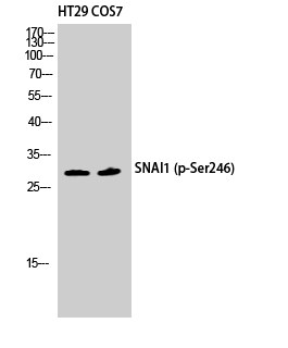
SNAI 1 (phospho Ser246) Polyclonal Antibody
- 产品详情
- 实验流程
- 背景知识
Application
| WB, IHC-P, IF |
|---|---|
| Primary Accession | O95863 |
| Reactivity | Human, Mouse, Monkey |
| Host | Rabbit |
| Clonality | Polyclonal |
| Calculated MW | 29083 Da |
| Gene ID | 6615 |
|---|---|
| Other Names | SNAI1; SNAH; Zinc finger protein SNAI1; Protein snail homolog 1; Protein sna |
| Dilution | WB~~Western Blot: 1/500 - 1/2000. Immunohistochemistry: 1/100 - 1/300. Immunofluorescence: 1/200 - 1/1000. ELISA: 1/5000. Not yet tested in other applications. IHC-P~~N/A IF~~1:50~200 |
| Format | Liquid in PBS containing 50% glycerol, 0.5% BSA and 0.09% (W/V) sodium azide. |
| Storage Conditions | -20℃ |
| Name | SNAI1 |
|---|---|
| Synonyms | SNAH |
| Function | Involved in induction of the epithelial to mesenchymal transition (EMT), formation and maintenance of embryonic mesoderm, growth arrest, survival and cell migration (PubMed:10655587, PubMed:15647282, PubMed:20389281, PubMed:20562920, PubMed:21952048, PubMed:25827072). Binds to 3 E-boxes of the E-cadherin/CDH1 gene promoter and to the promoters of CLDN7 and KRT8 and, in association with histone demethylase KDM1A which it recruits to the promoters, causes a decrease in dimethylated H3K4 levels and represses transcription (PubMed:10655587, PubMed:20389281, PubMed:20562920). The N-terminal SNAG domain competes with histone H3 for the same binding site on the histone demethylase complex formed by KDM1A and RCOR1, and thereby inhibits demethylation of histone H3 at 'Lys-4' (in vitro) (PubMed:20389281, PubMed:21300290, PubMed:23721412). During EMT, involved with LOXL2 in negatively regulating pericentromeric heterochromatin transcription (PubMed:16096638). SNAI1 recruits LOXL2 to pericentromeric regions to oxidize histone H3 and repress transcription which leads to release of heterochromatin component CBX5/HP1A, enabling chromatin reorganization and acquisition of mesenchymal traits (By similarity). Associates with EGR1 and SP1 to mediate tetradecanoyl phorbol acetate (TPA)-induced up-regulation of CDKN2B, possibly by binding to the CDKN2B promoter region 5'-TCACA-3 (PubMed:20121949). In addition, may also activate the CDKN2B promoter by itself (PubMed:20121949). |
| Cellular Location | Nucleus. Cytoplasm. Note=Once phosphorylated (probably on Ser-107, Ser-111, Ser-115 and Ser-119) it is exported from the nucleus to the cytoplasm where subsequent phosphorylation of the destruction motif and ubiquitination involving BTRC occurs. |
| Tissue Location | Expressed in a variety of tissues with the highest expression in kidney. Expressed in mesenchymal and epithelial cell lines. |
For Research Use Only. Not For Use In Diagnostic Procedures.
Provided below are standard protocols that you may find useful for product applications.
BACKGROUND
Involved in induction of the epithelial to mesenchymal transition (EMT), formation and maintenance of embryonic mesoderm, growth arrest, survival and cell migration. Binds to 3 E-boxes of the E-cadherin/CDH1 gene promoter and to the promoters of CLDN7 and KRT8 and, in association with histone demethylase KDM1A which it recruits to the promoters, causes a decrease in dimethylated H3K4 levels and represses transcription. During EMT, involved with LOXL2 in negatively regulating pericentromeric heterochromatin transcription (By similarity). SNAI1 recruits LOXL2 to pericentromeric regions to oxidize histone H3 and repress transcription which leads to release of heterochromatin component CBX5/HP1A, enabling chromatin reorganization and acquisition of mesenchymal traits (By similarity). Associates with EGR1 and SP1 to mediate tetradecanoyl phorbol acetate (TPA)-induced up- regulation of CDKN2B, possibly by binding to the CDKN2B promoter region 5'-TCACA-3. In addition, may also activate the CDKN2B promoter by itself.
终于等到您。ABCEPTA(百远生物)抗体产品。
点击下方“我要评价 ”按钮提交您的反馈信息,您的反馈和评价是我们最宝贵的财富之一,
我们将在1-3个工作日内处理您的反馈信息。
如有疑问,联系:0512-88856768 tech-china@abcepta.com.


