


 癌症的基本特征包括细胞增殖、血管生成、迁移、凋亡逃避机制和细胞永生等。找到癌症发生过程中这些通路的关键标记物和对应的抗体用于检测至关重要。
 为您推荐一个泛素化位点预测神器——泛素化分析工具,可以为您的蛋白的泛素化位点作出预测和评分。
 细胞自噬受体图形绘图工具为你的蛋白的细胞受体结合位点作出预测和评分,识别结合到自噬通路中的蛋白是非常重要的,便于让我们理解自噬在正常生理、病理过程中的作用,如发育、细胞分化、神经退化性疾病、压力条件下、感染和癌症。
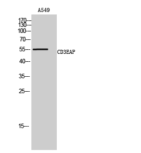
CD3EAP Polyclonal Antibody
- 产品详情
- 实验流程
- 背景知识
Application
| WB, IHC-P |
|---|---|
| Primary Accession | O15446 |
| Reactivity | Human |
| Host | Rabbit |
| Clonality | Polyclonal |
| Calculated MW | 54986 Da |
| Gene ID | 10849 |
|---|---|
| Other Names | CD3EAP; ASE1; CAST; PAF49; DNA-directed RNA polymerase I subunit RPA34; A34.5; Antisense to ERCC-1 protein; ASE-1; CD3-epsilon-associated protein; CAST; CD3E-associated protein; RNA polymerase I-associated factor PAF49 |
| Dilution | WB~~Western Blot: 1/500 - 1/2000. Immunohistochemistry: 1/100 - 1/300. ELISA: 1/40000. Not yet tested in other applications. IHC-P~~N/A |
| Format | Liquid in PBS containing 50% glycerol, 0.5% BSA and 0.09% (W/V) sodium azide. |
| Storage Conditions | -20℃ |
| Name | POLR1G (HGNC:24219) |
|---|---|
| Function | Component of RNA polymerase I (Pol I), a DNA-dependent RNA polymerase which synthesizes ribosomal RNA precursors using the four ribonucleoside triphosphates as substrates. Involved in UBTF-activated transcription, presumably at a step following PIC formation. |
| Cellular Location | Nucleus, nucleolus. Chromosome. Note=Found at the fibrillar centers of the nucleolus in interphase and during cell division it is localized to the nucleolus organizer regions of the chromosomes |
Research Areas
For Research Use Only. Not For Use In Diagnostic Procedures.
Application Protocols
Provided below are standard protocols that you may find useful for product applications.
BACKGROUND
DNA-dependent RNA polymerase catalyzes the transcription of DNA into RNA using the four ribonucleoside triphosphates as substrates. Component of RNA polymerase I which synthesizes ribosomal RNA precursors. Isoform 1 is involved in UBTF-activated transcription, presumably at a step following PIC formation.
终于等到您。ABCEPTA(百远生物)抗体产品。
点击下方“我要评价 ”按钮提交您的反馈信息,您的反馈和评价是我们最宝贵的财富之一,
我们将在1-3个工作日内处理您的反馈信息。
如有疑问,联系:0512-88856768 tech-china@abcepta.com.


