


 癌症的基本特征包括细胞增殖、血管生成、迁移、凋亡逃避机制和细胞永生等。找到癌症发生过程中这些通路的关键标记物和对应的抗体用于检测至关重要。
 为您推荐一个泛素化位点预测神器——泛素化分析工具,可以为您的蛋白的泛素化位点作出预测和评分。
 细胞自噬受体图形绘图工具为你的蛋白的细胞受体结合位点作出预测和评分,识别结合到自噬通路中的蛋白是非常重要的,便于让我们理解自噬在正常生理、病理过程中的作用,如发育、细胞分化、神经退化性疾病、压力条件下、感染和癌症。
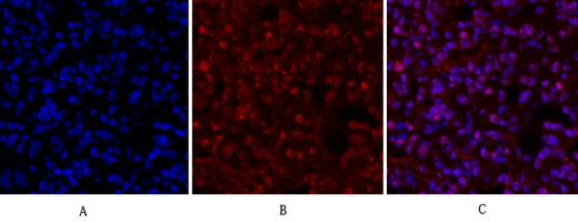
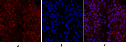
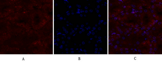
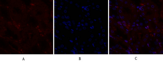
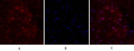
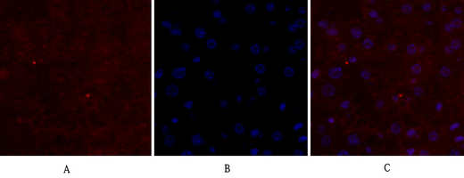
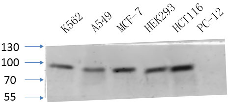
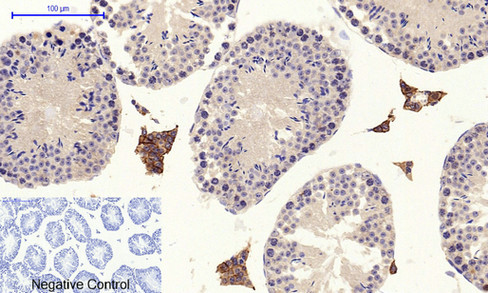
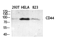
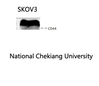
CD44 Polyclonal Antibody
.jpg)
- 产品详情
- 实验流程
- 背景知识
Application
| WB, IHC-P, IF |
|---|---|
| Primary Accession | P16070 |
| Reactivity | Human, Mouse, Rat |
| Host | Rabbit |
| Clonality | Polyclonal |
| Calculated MW | 81538 Da |
| Gene ID | 960 |
|---|---|
| Other Names | CD44; LHR; MDU2; MDU3; MIC4; CD44 antigen; CDw44; Epican; Extracellular matrix receptor III; ECMR-III; GP90 lymphocyte homing/adhesion receptor; HUTCH-I; Heparan sulfate proteoglycan; Hermes antigen; Hyaluronate receptor; Phagocytic glycopr |
| Dilution | WB~~IF: 1:50-200 WB 1:500-2000, IHC 1:50-300 IHC-P~~IF: 1:50-200 WB 1:500-2000, IHC 1:50-300 IF~~IF: 1:50-200 WB 1:500-2000, IHC 1:50-300 |
| Format | Liquid in PBS containing 50% glycerol, 0.5% BSA and 0.09% (W/V) sodium azide. |
| Storage Conditions | -20℃ |
| Name | CD44 |
|---|---|
| Synonyms | LHR, MDU2, MDU3, MIC4 |
| Function | Cell-surface receptor that plays a role in cell-cell interactions, cell adhesion and migration, helping them to sense and respond to changes in the tissue microenvironment (PubMed:16541107, PubMed:19703720, PubMed:22726066). Participates thereby in a wide variety of cellular functions including the activation, recirculation and homing of T-lymphocytes, hematopoiesis, inflammation and response to bacterial infection (PubMed:7528188). Engages, through its ectodomain, extracellular matrix components such as hyaluronan/HA, collagen, growth factors, cytokines or proteases and serves as a platform for signal transduction by assembling, via its cytoplasmic domain, protein complexes containing receptor kinases and membrane proteases (PubMed:18757307, PubMed:23589287). Such effectors include PKN2, the RhoGTPases RAC1 and RHOA, Rho-kinases and phospholipase C that coordinate signaling pathways promoting calcium mobilization and actin-mediated cytoskeleton reorganization essential for cell migration and adhesion (PubMed:15123640). |
| Cellular Location | Cell membrane; Single-pass type I membrane protein. Cell projection, microvillus {ECO:0000250|UniProtKB:P15379}. Secreted Note=Colocalizes with actin in membrane protrusions at wounding edges Co-localizes with RDX, EZR and MSN in microvilli. Localizes to cholesterol-rich membrane-bound lipid raft domains {ECO:0000250|UniProtKB:P15379, ECO:0000269|PubMed:23589287} |
| Tissue Location | Detected in fibroblasts and urine (at protein level) (PubMed:25326458, PubMed:36213313, PubMed:37453717). Detected in placenta (at protein level) (PubMed:32337544). Isoform 10 (epithelial isoform) is expressed by cells of epithelium and highly expressed by carcinomas. Expression is repressed in neuroblastoma cells |
For Research Use Only. Not For Use In Diagnostic Procedures.
Provided below are standard protocols that you may find useful for product applications.
BACKGROUND
Receptor for hyaluronic acid (HA). Mediates cell-cell and cell-matrix interactions through its affinity for HA, and possibly also through its affinity for other ligands such as osteopontin, collagens, and matrix metalloproteinases (MMPs). Adhesion with HA plays an important role in cell migration, tumor growth and progression. In cancer cells, may play an important role in invadopodia formation. Also involved in lymphocyte activation, recirculation and homing, and in hematopoiesis. Altered expression or dysfunction causes numerous pathogenic phenotypes. Great protein heterogeneity due to numerous alternative splicing and post-translational modification events. Receptor for LGALS9; the interaction enhances binding of SMAD3 to the FOXP3 promoter, leading to up-regulation of FOXP3 expression and increased induced regulatory T (iTreg) cell stability and suppressive function (By similarity).
终于等到您。ABCEPTA(百远生物)抗体产品。
点击下方“我要评价 ”按钮提交您的反馈信息,您的反馈和评价是我们最宝贵的财富之一,
我们将在1-3个工作日内处理您的反馈信息。
如有疑问,联系:0512-88856768 tech-china@abcepta.com.


