


 癌症的基本特征包括细胞增殖、血管生成、迁移、凋亡逃避机制和细胞永生等。找到癌症发生过程中这些通路的关键标记物和对应的抗体用于检测至关重要。
 为您推荐一个泛素化位点预测神器——泛素化分析工具,可以为您的蛋白的泛素化位点作出预测和评分。
 细胞自噬受体图形绘图工具为你的蛋白的细胞受体结合位点作出预测和评分,识别结合到自噬通路中的蛋白是非常重要的,便于让我们理解自噬在正常生理、病理过程中的作用,如发育、细胞分化、神经退化性疾病、压力条件下、感染和癌症。
NDRG1 Antibody (N-term)
Affinity Purified Rabbit Polyclonal Antibody (Pab)
- 产品详情
- 文献引用 : 2
- 实验流程
- 背景知识
Application
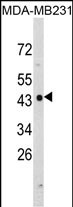
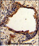
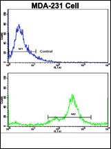
| WB, IHC-P, FC, E |
|---|---|
| Primary Accession | Q92597 |
| Other Accession | Q6JE36, Q62433, Q4R4Q3, Q3SYX0 |
| Reactivity | Human |
| Predicted | Bovine, Monkey, Mouse, Rat |
| Host | Rabbit |
| Clonality | Polyclonal |
| Isotype | Rabbit IgG |
| Calculated MW | 42835 Da |
| Antigen Region | 12-40 aa |
| Gene ID | 10397 |
|---|---|
| Other Names | Protein NDRG1, Differentiation-related gene 1 protein, DRG-1, N-myc downstream-regulated gene 1 protein, Nickel-specific induction protein Cap43, Reducing agents and tunicamycin-responsive protein, RTP, Rit42, NDRG1, CAP43, DRG1, RTP |
| Target/Specificity | This NDRG1 antibody is generated from rabbits immunized with a KLH conjugated synthetic peptide between 12-40 amino acids from the N-terminal region of human NDRG1. |
| Dilution | WB~~1:1000 IHC-P~~1:100~500 FC~~1:10~50 E~~Use at an assay dependent concentration. |
| Format | Purified polyclonal antibody supplied in PBS with 0.09% (W/V) sodium azide. This antibody is purified through a protein A column, followed by peptide affinity purification. |
| Storage | Maintain refrigerated at 2-8°C for up to 2 weeks. For long term storage store at -20°C in small aliquots to prevent freeze-thaw cycles. |
| Precautions | NDRG1 Antibody (N-term) is for research use only and not for use in diagnostic or therapeutic procedures. |
| Name | NDRG1 |
|---|---|
| Synonyms | CAP43, DRG1, RTP |
| Function | Stress-responsive protein involved in hormone responses, cell growth, and differentiation. Acts as a tumor suppressor in many cell types. Necessary but not sufficient for p53/TP53-mediated caspase activation and apoptosis. Has a role in cell trafficking, notably of the Schwann cell, and is necessary for the maintenance and development of the peripheral nerve myelin sheath. Required for vesicular recycling of CDH1 and TF. May also function in lipid trafficking. Protects cells from spindle disruption damage. Functions in p53/TP53-dependent mitotic spindle checkpoint. Regulates microtubule dynamics and maintains euploidy. |
| Cellular Location | Cytoplasm, cytosol. Cytoplasm, cytoskeleton, microtubule organizing center, centrosome. Nucleus. Cell membrane Note=Mainly cytoplasmic but differentially localized to other regions Associates with the plasma membrane in intestinal epithelia and lactating mammary gland. Translocated to the nucleus in a p53/TP53- dependent manner. In prostate epithelium and placental chorion, located in both the cytoplasm and in the nucleus. No nuclear localization in colon epithelium cells. In intestinal mucosa, prostate and renal cortex, located predominantly adjacent to adherens junctions Cytoplasmic with granular staining in proximal tubular cells of the kidney and salivary gland ducts. Recruits to the membrane of recycling/sorting and late endosomes via binding to phosphatidylinositol 4-phosphate. Associates with microtubules Colocalizes with TUBG1 in the centrosome. Cytoplasmic location increased with hypoxia. Phosphorylated form found associated with centromeres during S-phase of mitosis and with the plasma membrane |
| Tissue Location | Ubiquitous; expressed most prominently in placental membranes and prostate, kidney, small intestine, and ovary tissues Also expressed in heart, brain, skeletal muscle, lung, liver and pancreas. Low levels in peripheral blood leukocytes and in tissues of the immune system. Expressed mainly in epithelial cells. Also found in Schwann cells of peripheral neurons. Reduced expression in adenocarcinomas compared to normal tissues. In colon, prostate and placental membranes, the cells that border the lumen show the highest expression. |
Research Areas
For Research Use Only. Not For Use In Diagnostic Procedures.

Application Protocols
Provided below are standard protocols that you may find useful for product applications.
BACKGROUND
NDRG1 is a cytoplasmic protein involved in stress responses, hormone responses, cell growth, and differentiation. It is necessary for p53-mediated caspase activation and apoptosis.
REFERENCES
Sugiyama,N., et.al., Mol. Cell Proteomics 6 (6), 1103-1109 (2007)
终于等到您。ABCEPTA(百远生物)抗体产品。
点击下方“我要评价 ”按钮提交您的反馈信息,您的反馈和评价是我们最宝贵的财富之一,
我们将在1-3个工作日内处理您的反馈信息。
如有疑问,联系:0512-88856768 tech-china@abcepta.com.
¥ 1,250.00
Cat# AP6935a


