


 癌症的基本特征包括细胞增殖、血管生成、迁移、凋亡逃避机制和细胞永生等。找到癌症发生过程中这些通路的关键标记物和对应的抗体用于检测至关重要。
 为您推荐一个泛素化位点预测神器——泛素化分析工具,可以为您的蛋白的泛素化位点作出预测和评分。
 细胞自噬受体图形绘图工具为你的蛋白的细胞受体结合位点作出预测和评分,识别结合到自噬通路中的蛋白是非常重要的,便于让我们理解自噬在正常生理、病理过程中的作用,如发育、细胞分化、神经退化性疾病、压力条件下、感染和癌症。
eIF1AY Polyclonal Antibody
- 产品详情
- 实验流程
- 背景知识
Application
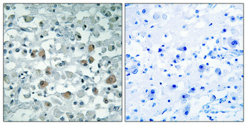
| IHC-P, IF, ICC, E |
|---|---|
| Primary Accession | O14602 |
| Reactivity | Human, Mouse, Rat |
| Host | Rabbit |
| Clonality | Polyclonal |
| Calculated MW | 16442 Da |
| Gene ID | 9086 |
|---|---|
| Other Names | EIF1AY; Eukaryotic translation initiation factor 1A; Y-chromosomal; eIF-1A Y isoform; Eukaryotic translation initiation factor 4C; eIF-4C |
| Dilution | IHC-P~~Immunohistochemistry: 1/100 - 1/300. ELISA: 1/40000. Not yet tested in other applications. IF~~1:50~200 ICC~~N/A E~~N/A |
| Format | Liquid in PBS containing 50% glycerol, 0.5% BSA and 0.09% (W/V) sodium azide. |
| Storage Conditions | -20℃ |
| Name | EIF1AY |
|---|---|
| Function | Component of the 43S pre-initiation complex (43S PIC), which binds to the mRNA cap-proximal region, scans mRNA 5'-untranslated region, and locates the initiation codon. This protein enhances formation of the cap-proximal complex. Together with EIF1, facilitates scanning, start codon recognition, promotion of the assembly of 48S complex at the initiation codon (43S PIC becomes 48S PIC after the start codon is reached), and dissociation of aberrant complexes. After start codon location, together with EIF5B orients the initiator methionine-tRNA in a conformation that allows 60S ribosomal subunit joining to form the 80S initiation complex. Is released after 80S initiation complex formation, just after GTP hydrolysis by EIF5B, and before release of EIF5B. Its globular part is located in the A site of the 40S ribosomal subunit. Its interaction with EIF5 during scanning contribute to the maintenance of EIF1 within the open 43S PIC. In contrast to yeast orthologs, does not bind EIF1. |
| Cellular Location | Cytoplasm. |
| Tissue Location | Ubiquitous. |
Research Areas
For Research Use Only. Not For Use In Diagnostic Procedures.
Application Protocols
Provided below are standard protocols that you may find useful for product applications.
BACKGROUND
Seems to be required for maximal rate of protein biosynthesis. Enhances ribosome dissociation into subunits and stabilizes the binding of the initiator Met-tRNA(I) to 40 S ribosomal subunits (By similarity).
终于等到您。ABCEPTA(百远生物)抗体产品。
点击下方“我要评价 ”按钮提交您的反馈信息,您的反馈和评价是我们最宝贵的财富之一,
我们将在1-3个工作日内处理您的反馈信息。
如有疑问,联系:0512-88856768 tech-china@abcepta.com.
¥ 1,500.00
Cat# AP69686


