


 癌症的基本特征包括细胞增殖、血管生成、迁移、凋亡逃避机制和细胞永生等。找到癌症发生过程中这些通路的关键标记物和对应的抗体用于检测至关重要。
 为您推荐一个泛素化位点预测神器——泛素化分析工具,可以为您的蛋白的泛素化位点作出预测和评分。
 细胞自噬受体图形绘图工具为你的蛋白的细胞受体结合位点作出预测和评分,识别结合到自噬通路中的蛋白是非常重要的,便于让我们理解自噬在正常生理、病理过程中的作用,如发育、细胞分化、神经退化性疾病、压力条件下、感染和癌症。
LAG3 Antibody (Center)
Affinity Purified Rabbit Polyclonal Antibody (Pab)
- 产品详情
- 实验流程
- 背景知识
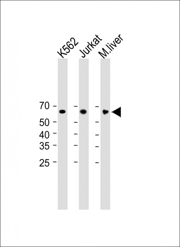
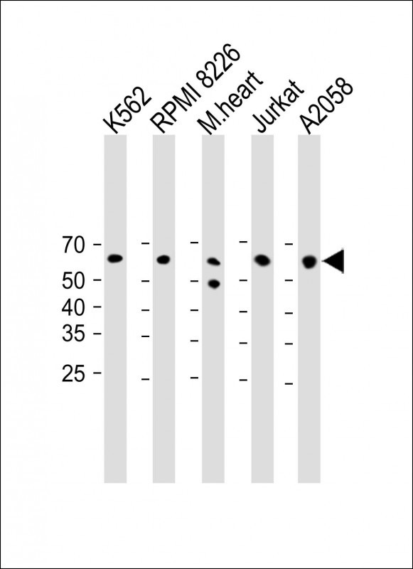
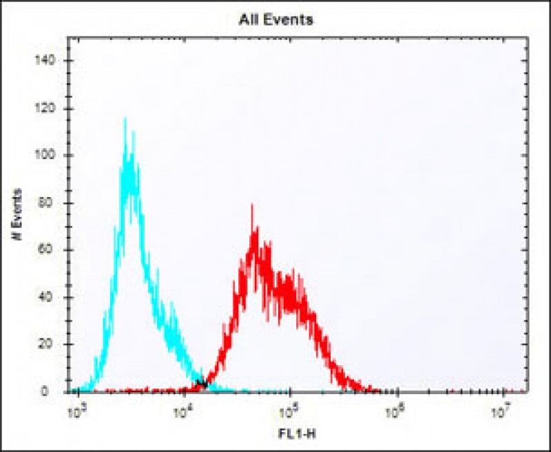
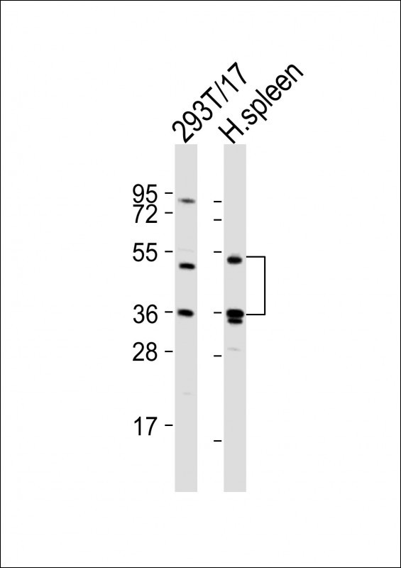
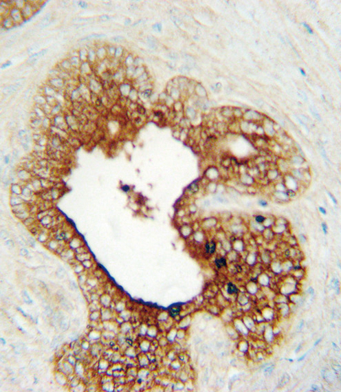
Application
| WB, IHC-P, FC, E |
|---|---|
| Primary Accession | P18627 |
| Reactivity | Human, Mouse, Rat |
| Host | Rabbit |
| Clonality | Polyclonal |
| Isotype | Rabbit IgG |
| Calculated MW | 57449 Da |
| Antigen Region | 103-132 aa |
| Gene ID | 3902 |
|---|---|
| Other Names | Lymphocyte activation gene 3 protein, LAG-3, Protein FDC, CD223, LAG3, FDC |
| Target/Specificity | This LAG3 antibody is generated from rabbits immunized with a KLH conjugated synthetic peptide between 103-132 amino acids from the Central region of human LAG3. |
| Dilution | WB~~1:2000 IHC-P~~1:100~500 FC~~1:25 E~~Use at an assay dependent concentration. |
| Format | Purified polyclonal antibody supplied in PBS with 0.09% (W/V) sodium azide. This antibody is purified through a protein A column, followed by peptide affinity purification. |
| Storage | Maintain refrigerated at 2-8°C for up to 2 weeks. For long term storage store at -20°C in small aliquots to prevent freeze-thaw cycles. |
| Precautions | LAG3 Antibody (Center) is for research use only and not for use in diagnostic or therapeutic procedures. |
| Name | LAG3 {ECO:0000303|PubMed:35761082, ECO:0000312|HGNC:HGNC:6476} |
|---|---|
| Function | [Lymphocyte activation gene 3 protein]: Inhibitory receptor on antigen activated T-cells (PubMed:20421648, PubMed:35761082, PubMed:7805750, PubMed:8647185). Delivers inhibitory signals upon binding to ligands, such as MHC class II, its main ligand present at the surface of antigen-presenting cells (APCs), and FGL1, which is secreted by hepatocytes and certain types of tumor cells (PubMed:30580966, PubMed:32920841, PubMed:35761082, PubMed:39671469, PubMed:7589152, PubMed:8647185, PubMed:9159144). Ligand-binding initiates a signaling that inhibits the T-cell receptor (TCR) in the immunological synapse, preventing T-cell activation (PubMed:40101708). Mechanistically, ligand-binding promotes (1) ubiquitination of the KIEELE motif, unleashing the RRFSALE motif from the membrane and (2) leading to the formation of condensates with the TCR component CD3E, thereby disrupting the association between CD3E and LCK and preventing TCR activation (PubMed:40101708, PubMed:40592325). May inhibit antigen- specific T-cell activation in synergy with PDCD1/PD-1 (By similarity). Negatively regulates the proliferation, activation, effector function and homeostasis of both CD8(+) and CD4(+) T-cells (PubMed:20421648, PubMed:7805750, PubMed:8647185). Also mediates immune tolerance: constitutively expressed on a subset of regulatory T-cells (Tregs) and contributes to their suppressive function (By similarity). Also acts as a negative regulator of plasmacytoid dendritic cell (pDCs) activation (By similarity). |
| Cellular Location | [Lymphocyte activation gene 3 protein]: Cell membrane; Single-pass type I membrane protein. Note=Clusters on the T-cell surface following ligand-binding |
| Tissue Location | Primarily expressed in activated T-cells and a subset of natural killer (NK) cells. |
Research Areas
For Research Use Only. Not For Use In Diagnostic Procedures.
Application Protocols
Provided below are standard protocols that you may find useful for product applications.
BACKGROUND
Lymphocyte-activation protein 3 belongs to Ig superfamily and contains 4 extracellular Ig-like domains.
REFERENCES
Smyth,D.J., et.al., BMC Med. Genet. 7, 20 (2006)
终于等到您。ABCEPTA(百远生物)抗体产品。
点击下方“我要评价 ”按钮提交您的反馈信息,您的反馈和评价是我们最宝贵的财富之一,
我们将在1-3个工作日内处理您的反馈信息。
如有疑问,联系:0512-88856768 tech-china@abcepta.com.
¥ 699.00
Cat# AP6987c


