


 癌症的基本特征包括细胞增殖、血管生成、迁移、凋亡逃避机制和细胞永生等。找到癌症发生过程中这些通路的关键标记物和对应的抗体用于检测至关重要。
 为您推荐一个泛素化位点预测神器——泛素化分析工具,可以为您的蛋白的泛素化位点作出预测和评分。
 细胞自噬受体图形绘图工具为你的蛋白的细胞受体结合位点作出预测和评分,识别结合到自噬通路中的蛋白是非常重要的,便于让我们理解自噬在正常生理、病理过程中的作用,如发育、细胞分化、神经退化性疾病、压力条件下、感染和癌症。
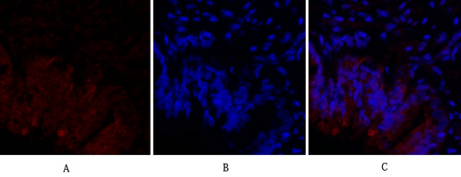
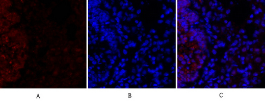
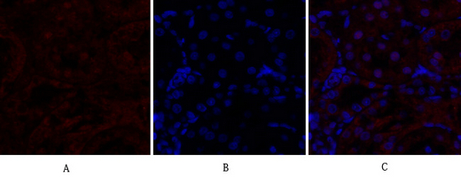
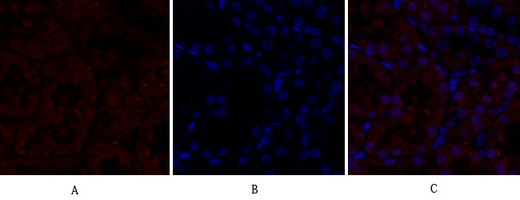
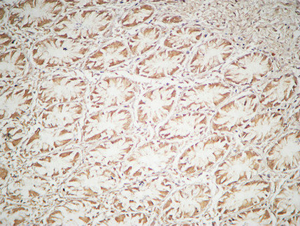
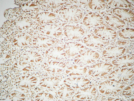
HIF-1α Polyclonal Antibody
- 产品详情
- 实验流程
- 背景知识
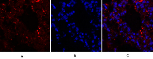
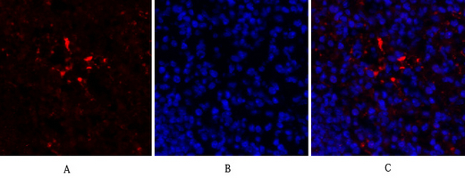
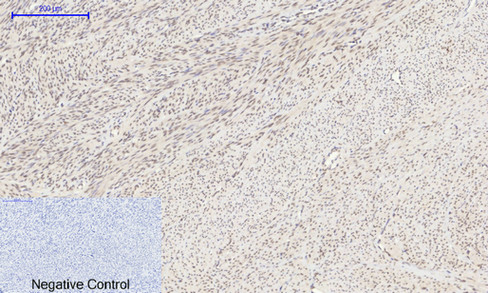
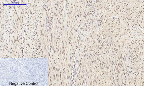
Application
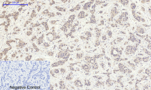
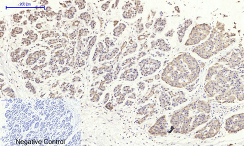
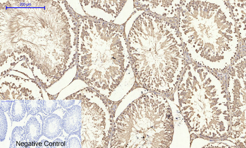
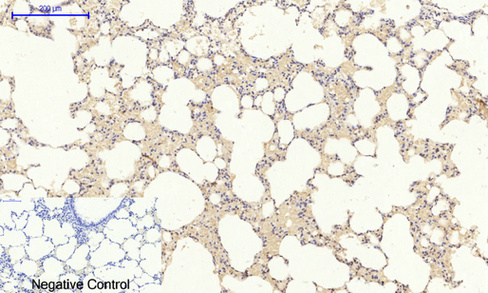
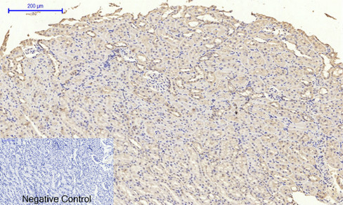
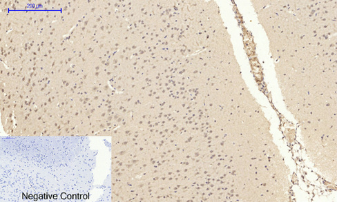
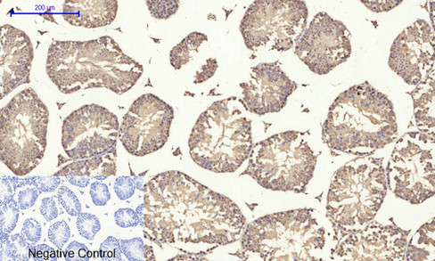
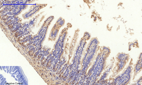
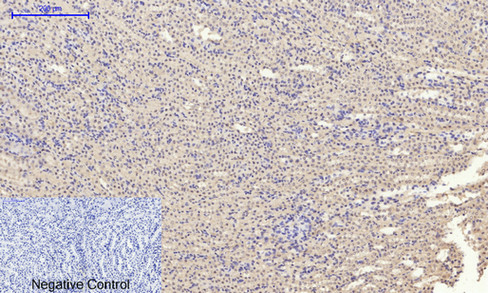
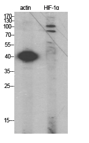
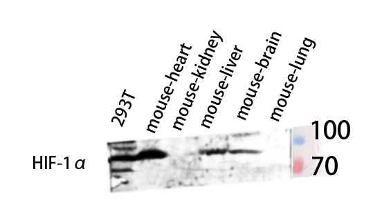
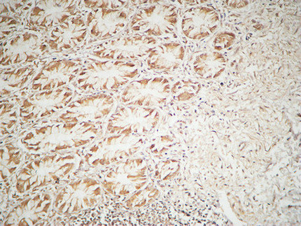
| IF, ICC, WB, IHC-P, IP, E |
|---|---|
| Primary Accession | Q16665 |
| Reactivity | Human, Mouse, Rat |
| Host | Rabbit |
| Clonality | Polyclonal |
| Calculated MW | 92670 Da |
| Gene ID | 3091 |
|---|---|
| Other Names | HIF1A; BHLHE78; MOP1; PASD8; Hypoxia-inducible factor 1-alpha; HIF-1-alpha; HIF1-alpha; ARNT-interacting protein; Basic-helix-loop-helix-PAS protein MOP1; Class E basic helix-loop-helix protein 78; bHLHe78; Member of PAS protein 1; PAS doma |
| Dilution | IF~~IF: 1:50-200 Western Blot: 1/500 - 1/2000. Immunohistochemistry: 1/100 - 1/300. ELISA: 1/40000. Not yet tested in other applications. ICC~~N/A WB~~IF: 1:50-200 Western Blot: 1/500 - 1/2000. Immunohistochemistry: 1/100 - 1/300. ELISA: 1/40000. Not yet tested in other applications. IHC-P~~IF: 1:50-200 Western Blot: 1/500 - 1/2000. Immunohistochemistry: 1/100 - 1/300. ELISA: 1/40000. Not yet tested in other applications. IP~~N/A E~~N/A |
| Format | Liquid in PBS containing 50% glycerol, 0.5% BSA and 0.09% (W/V) sodium azide. |
| Storage Conditions | -20℃ |
| Name | HIF1A {ECO:0000303|PubMed:7539918, ECO:0000312|HGNC:HGNC:4910} |
|---|---|
| Function | Functions as a master transcriptional regulator of the adaptive response to hypoxia (PubMed:11292861, PubMed:11566883, PubMed:15465032, PubMed:16973622, PubMed:17610843, PubMed:18658046, PubMed:20624928, PubMed:22009797, PubMed:30125331, PubMed:9887100). Under hypoxic conditions, activates the transcription of over 40 genes, including erythropoietin, glucose transporters, glycolytic enzymes, vascular endothelial growth factor, HILPDA, and other genes whose protein products increase oxygen delivery or facilitate metabolic adaptation to hypoxia (PubMed:11292861, PubMed:11566883, PubMed:15465032, PubMed:16973622, PubMed:17610843, PubMed:20624928, PubMed:22009797, PubMed:30125331, PubMed:9887100). Plays an essential role in embryonic vascularization, tumor angiogenesis and pathophysiology of ischemic disease (PubMed:22009797). Heterodimerizes with ARNT; heterodimer binds to core DNA sequence 5'-TACGTG-3' within the hypoxia response element (HRE) of target gene promoters (By similarity). Activation requires recruitment of transcriptional coactivators such as CREBBP and EP300 (PubMed:16543236, PubMed:9887100). Activity is enhanced by interaction with NCOA1 and/or NCOA2 (PubMed:10594042). Interaction with redox regulatory protein APEX1 seems to activate CTAD and potentiates activation by NCOA1 and CREBBP (PubMed:10202154, PubMed:10594042). Involved in the axonal distribution and transport of mitochondria in neurons during hypoxia (PubMed:19528298). |
| Cellular Location | Cytoplasm. Nucleus. Nucleus speckle {ECO:0000250|UniProtKB:Q61221}. Note=Colocalizes with HIF3A in the nucleus and speckles (By similarity). Cytoplasmic in normoxia, nuclear translocation in response to hypoxia (PubMed:9822602) {ECO:0000250|UniProtKB:Q61221, ECO:0000269|PubMed:9822602} |
| Tissue Location | Expressed in most tissues with highest levels in kidney and heart. Overexpressed in the majority of common human cancers and their metastases, due to the presence of intratumoral hypoxia and as a result of mutations in genes encoding oncoproteins and tumor suppressors. A higher level expression seen in pituitary tumors as compared to the pituitary gland. |
For Research Use Only. Not For Use In Diagnostic Procedures.
Provided below are standard protocols that you may find useful for product applications.
BACKGROUND
Functions as a master transcriptional regulator of the adaptive response to hypoxia. Under hypoxic conditions, activates the transcription of over 40 genes, including erythropoietin, glucose transporters, glycolytic enzymes, vascular endothelial growth factor, HILPDA, and other genes whose protein products increase oxygen delivery or facilitate metabolic adaptation to hypoxia. Plays an essential role in embryonic vascularization, tumor angiogenesis and pathophysiology of ischemic disease. Heterodimerizes with ARNT; heterodimer binds to core DNA sequence 5'-TACGTG-3' within the hypoxia response element (HRE) of target gene promoters (By similarity). Activation requires recruitment of transcriptional coactivators such as CREBBP and EP300. Activity is enhanced by interaction with both, NCOA1 or NCOA2. Interaction with redox regulatory protein APEX seems to activate CTAD and potentiates activation by NCOA1 and CREBBP. Involved in the axonal distribution and transport of mitochondria in neurons during hypoxia.
终于等到您。ABCEPTA(百远生物)抗体产品。
点击下方“我要评价 ”按钮提交您的反馈信息,您的反馈和评价是我们最宝贵的财富之一,
我们将在1-3个工作日内处理您的反馈信息。
如有疑问,联系:0512-88856768 tech-china@abcepta.com.


