


 癌症的基本特征包括细胞增殖、血管生成、迁移、凋亡逃避机制和细胞永生等。找到癌症发生过程中这些通路的关键标记物和对应的抗体用于检测至关重要。
 为您推荐一个泛素化位点预测神器——泛素化分析工具,可以为您的蛋白的泛素化位点作出预测和评分。
 细胞自噬受体图形绘图工具为你的蛋白的细胞受体结合位点作出预测和评分,识别结合到自噬通路中的蛋白是非常重要的,便于让我们理解自噬在正常生理、病理过程中的作用,如发育、细胞分化、神经退化性疾病、压力条件下、感染和癌症。
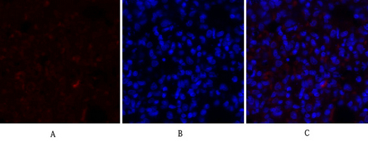
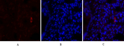
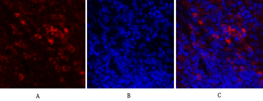
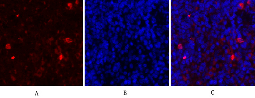
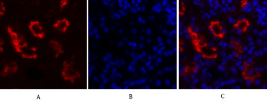
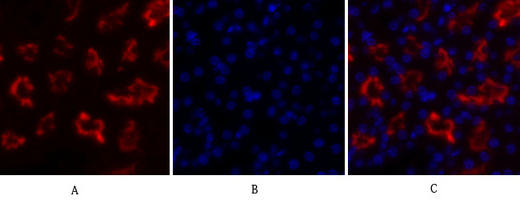
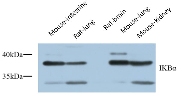
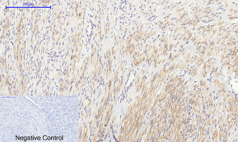
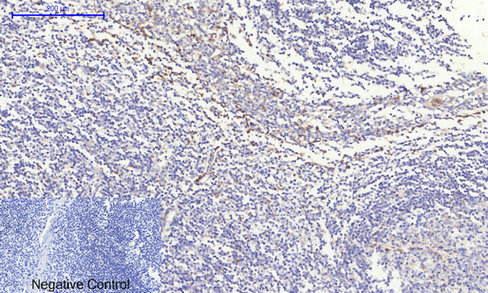
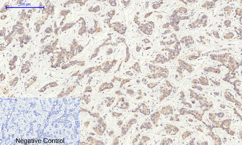
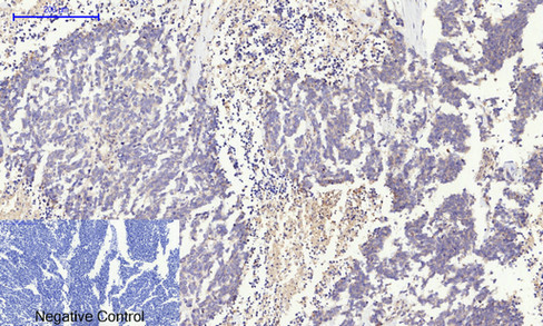
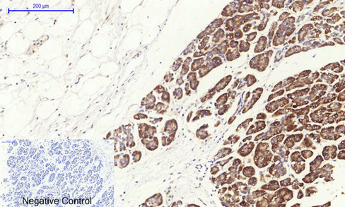
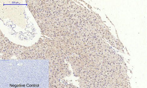
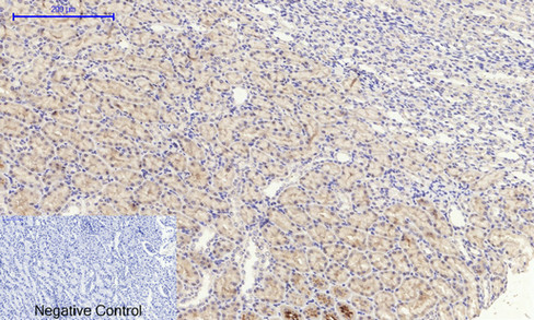
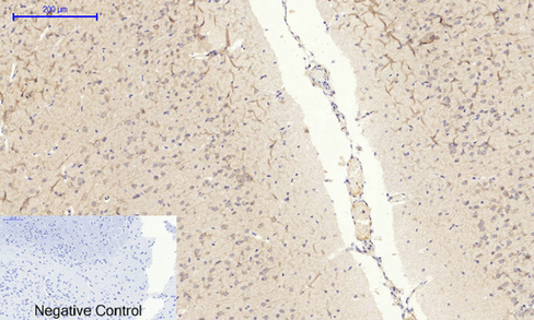
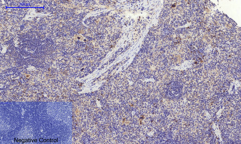
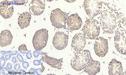
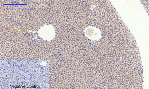
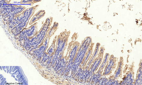
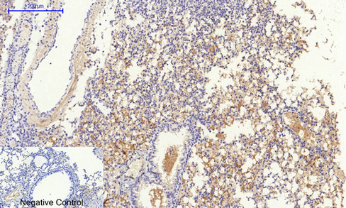
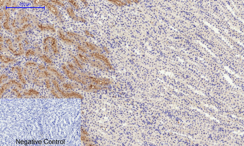
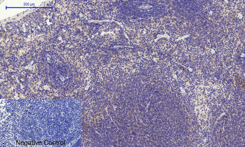
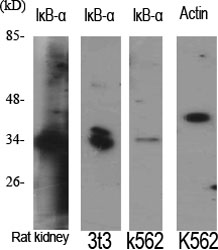
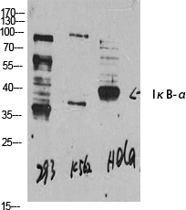
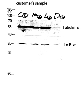
IκB-α Polyclonal Antibody
.jpg)
- 产品详情
- 实验流程
- 背景知识
Application
| WB, IHC-P, IF |
|---|---|
| Primary Accession | P25963 |
| Reactivity | Human, Mouse, Rat |
| Host | Rabbit |
| Clonality | Polyclonal |
| Calculated MW | 35609 Da |
| Gene ID | 4792 |
|---|---|
| Other Names | NFKBIA; IKBA; MAD3; NFKBI; NF-kappa-B inhibitor alpha; I-kappa-B-alpha; IkB-alpha; IkappaBalpha; Major histocompatibility complex enhancer-binding protein MAD3 |
| Dilution | WB~~IF: 1:50-200 WB 1:500-2000, ELISA 1:10000-20000 IHC 1:50-300 IHC-P~~IF: 1:50-200 WB 1:500-2000, ELISA 1:10000-20000 IHC 1:50-300 IF~~IF: 1:50-200 WB 1:500-2000, ELISA 1:10000-20000 IHC 1:50-300 |
| Format | Liquid in PBS containing 50% glycerol, 0.5% BSA and 0.09% (W/V) sodium azide. |
| Storage Conditions | -20℃ |
| Name | NFKBIA |
|---|---|
| Synonyms | IKBA, MAD3, NFKBI |
| Function | Inhibits the activity of dimeric NF-kappa-B/REL complexes by trapping REL (RELA/p65 and NFKB1/p50) dimers in the cytoplasm by masking their nuclear localization signals (PubMed:1493333, PubMed:36651806, PubMed:7479976). On cellular stimulation by immune and pro-inflammatory responses, becomes phosphorylated promoting ubiquitination and degradation, enabling the dimeric RELA to translocate to the nucleus and activate transcription (PubMed:7479976, PubMed:7628694, PubMed:7796813, PubMed:7878466). |
| Cellular Location | Cytoplasm. Nucleus. Note=Shuttles between the nucleus and the cytoplasm by a nuclear localization signal (NLS) and a CRM1-dependent nuclear export. |
For Research Use Only. Not For Use In Diagnostic Procedures.
Provided below are standard protocols that you may find useful for product applications.
BACKGROUND
Inhibits the activity of dimeric NF-kappa-B/REL complexes by trapping REL dimers in the cytoplasm through masking of their nuclear localization signals. On cellular stimulation by immune and proinflammatory responses, becomes phosphorylated promoting ubiquitination and degradation, enabling the dimeric RELA to translocate to the nucleus and activate transcription.
终于等到您。ABCEPTA(百远生物)抗体产品。
点击下方“我要评价 ”按钮提交您的反馈信息,您的反馈和评价是我们最宝贵的财富之一,
我们将在1-3个工作日内处理您的反馈信息。
如有疑问,联系:0512-88856768 tech-china@abcepta.com.


