


 癌症的基本特征包括细胞增殖、血管生成、迁移、凋亡逃避机制和细胞永生等。找到癌症发生过程中这些通路的关键标记物和对应的抗体用于检测至关重要。
 为您推荐一个泛素化位点预测神器——泛素化分析工具,可以为您的蛋白的泛素化位点作出预测和评分。
 细胞自噬受体图形绘图工具为你的蛋白的细胞受体结合位点作出预测和评分,识别结合到自噬通路中的蛋白是非常重要的,便于让我们理解自噬在正常生理、病理过程中的作用,如发育、细胞分化、神经退化性疾病、压力条件下、感染和癌症。
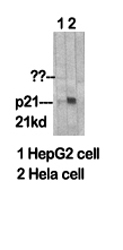
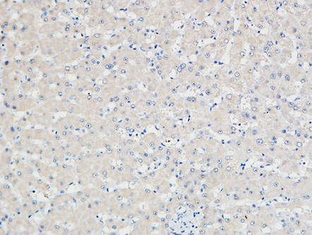
p21 Polyclonal Antibody
.jpg)
- 产品详情
- 实验流程
- 背景知识
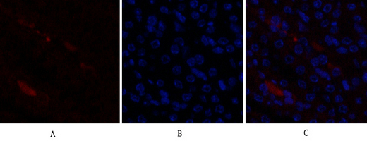
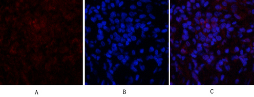
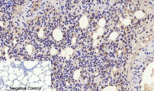
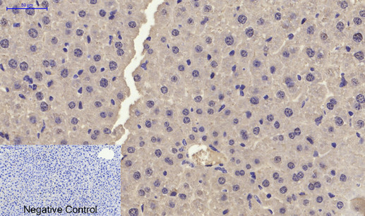
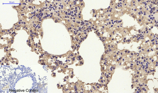
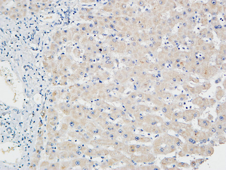
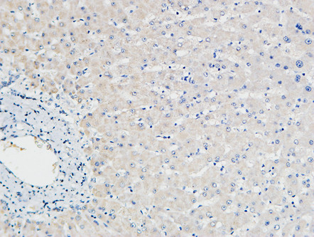
Application
| IF, ICC, WB, IHC-P, E |
|---|---|
| Primary Accession | P38936 |
| Reactivity | Human, Mouse, Rat |
| Host | Rabbit |
| Clonality | Polyclonal |
| Calculated MW | 18119 Da |
| Gene ID | 1026 |
|---|---|
| Other Names | CDKN1A; CAP20; CDKN1; CIP1; MDA6; PIC1; SDI1; WAF1; Cyclin-dependent kinase inhibitor 1; CDK-interacting protein 1; Melanoma differentiation-associated protein 6; MDA-6; p21 |
| Dilution | IF~~IF: 1:50-200 Western Blot: 1/500 - 1/2000. ELISA: 1/20000. Not yet tested in other applications. ICC~~N/A WB~~IF: 1:50-200 Western Blot: 1/500 - 1/2000. ELISA: 1/20000. Not yet tested in other applications. IHC-P~~IF: 1:50-200 Western Blot: 1/500 - 1/2000. ELISA: 1/20000. Not yet tested in other applications. E~~N/A |
| Format | Liquid in PBS containing 50% glycerol, 0.5% BSA and 0.09% (W/V) sodium azide. |
| Storage Conditions | -20℃ |
| Name | CDKN1A (HGNC:1784) |
|---|---|
| Function | Plays an important role in controlling cell cycle progression and DNA damage-induced G2 arrest (PubMed:9106657). Involved in p53/TP53 mediated inhibition of cellular proliferation in response to DNA damage. Also involved in p53-independent DNA damage-induced G2 arrest mediated by CREB3L1 in astrocytes and osteoblasts (By similarity). Binds to and inhibits cyclin-dependent kinase activity, preventing phosphorylation of critical cyclin-dependent kinase substrates and blocking cell cycle progression. Functions in the nuclear localization and assembly of cyclin D-CDK4 complex and promotes its kinase activity towards RB1. At higher stoichiometric ratios, inhibits the kinase activity of the cyclin D-CDK4 complex. Inhibits DNA synthesis by DNA polymerase delta by competing with POLD3 for PCNA binding (PubMed:11595739). Negatively regulates the CDK4- and CDK6-driven phosphorylation of RB1 in keratinocytes, thereby resulting in the release of E2F1 and subsequent transcription of E2F1-driven G1/S phase promoting genes (By similarity). |
| Cellular Location | Cytoplasm. Nucleus |
| Tissue Location | Expressed in all adult tissues, with 5-fold lower levels observed in the brain |
For Research Use Only. Not For Use In Diagnostic Procedures.
Provided below are standard protocols that you may find useful for product applications.
BACKGROUND
May be involved in p53/TP53 mediated inhibition of cellular proliferation in response to DNA damage. Binds to and inhibits cyclin-dependent kinase activity, preventing phosphorylation of critical cyclin-dependent kinase substrates and blocking cell cycle progression. Functions in the nuclear localization and assembly of cyclin D-CDK4 complex and promotes its kinase activity towards RB1. At higher stoichiometric ratios, inhibits the kinase activity of the cyclin D-CDK4 complex. Inhibits DNA synthesis by DNA polymerase delta by competing with POLD3 for PCNA binding (PubMed:11595739).
终于等到您。ABCEPTA(百远生物)抗体产品。
点击下方“我要评价 ”按钮提交您的反馈信息,您的反馈和评价是我们最宝贵的财富之一,
我们将在1-3个工作日内处理您的反馈信息。
如有疑问,联系:0512-88856768 tech-china@abcepta.com.


