


 癌症的基本特征包括细胞增殖、血管生成、迁移、凋亡逃避机制和细胞永生等。找到癌症发生过程中这些通路的关键标记物和对应的抗体用于检测至关重要。
 为您推荐一个泛素化位点预测神器——泛素化分析工具,可以为您的蛋白的泛素化位点作出预测和评分。
 细胞自噬受体图形绘图工具为你的蛋白的细胞受体结合位点作出预测和评分,识别结合到自噬通路中的蛋白是非常重要的,便于让我们理解自噬在正常生理、病理过程中的作用,如发育、细胞分化、神经退化性疾病、压力条件下、感染和癌症。
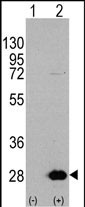
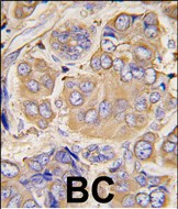
HSP27 (HSPB1) Antibody (S78)
Affinity Purified Rabbit Polyclonal Antibody (Pab)
- 产品详情
- 实验流程
- 背景知识
Application
| WB, IHC-P, E |
|---|---|
| Primary Accession | P04792 |
| Reactivity | Human |
| Host | Rabbit |
| Clonality | Polyclonal |
| Isotype | Rabbit IgG |
| Calculated MW | 22783 Da |
| Antigen Region | 56-85 aa |
| Gene ID | 3315 |
|---|---|
| Other Names | Heat shock protein beta-1, HspB1, 28 kDa heat shock protein, Estrogen-regulated 24 kDa protein, Heat shock 27 kDa protein, HSP 27, Stress-responsive protein 27, SRP27, HSPB1, HSP27, HSP28 |
| Target/Specificity | This HSP27(HSPB1) antibody is generated from rabbits immunized with a KLH conjugated synthetic peptide between 56-85 amino acids from human HSP27(HSPB1). |
| Dilution | WB~~1:1000 IHC-P~~1:100~500 E~~Use at an assay dependent concentration. |
| Format | Purified polyclonal antibody supplied in PBS with 0.09% (W/V) sodium azide. This antibody is purified through a protein A column, followed by peptide affinity purification. |
| Storage | Maintain refrigerated at 2-8°C for up to 2 weeks. For long term storage store at -20°C in small aliquots to prevent freeze-thaw cycles. |
| Precautions | HSP27 (HSPB1) Antibody (S78) is for research use only and not for use in diagnostic or therapeutic procedures. |
| Name | HSPB1 |
|---|---|
| Synonyms | HSP27, HSP28 |
| Function | Small heat shock protein which functions as a molecular chaperone probably maintaining denatured proteins in a folding- competent state (PubMed:10383393, PubMed:20178975). Plays a role in stress resistance and actin organization (PubMed:19166925). Through its molecular chaperone activity may regulate numerous biological processes including the phosphorylation and the axonal transport of neurofilament proteins (PubMed:23728742). |
| Cellular Location | Cytoplasm. Nucleus Cytoplasm, cytoskeleton, spindle Note=Cytoplasmic in interphase cells. Colocalizes with mitotic spindles in mitotic cells. Translocates to the nucleus during heat shock and resides in sub-nuclear structures known as SC35 speckles or nuclear splicing speckles. |
| Tissue Location | Detected in all tissues tested: skeletal muscle, heart, aorta, large intestine, small intestine, stomach, esophagus, bladder, adrenal gland, thyroid, pancreas, testis, adipose tissue, kidney, liver, spleen, cerebral cortex, blood serum and cerebrospinal fluid. Highest levels are found in the heart and in tissues composed of striated and smooth muscle. |
For Research Use Only. Not For Use In Diagnostic Procedures.
Provided below are standard protocols that you may find useful for product applications.
BACKGROUND
In response to adverse changes in their environment, cells from many organisms increase the expression of a class of proteins referred to as heat shock or stress proteins. HSBP1 exhibits rapid increased phosphorylation in response to various mitogens, tumor promoters (e.g. phorbol esters) and calcium ionophores, and high levels are associated with carcinoma of the breast and with endometrial adenocarcinomas. Heat shock of HeLa cell cultures, or treatment with arsenite, phorbol ester, or tumor necrosis factor, causes a rapid phosphorylation of preexisting HSBP1, with Ser82 as the major site and Ser78 the minor site of phosphorylation. HSBP1 may exert phosphorylation-activated functions linked with growth signaling pathways in unstressed cells. A homeostatic function at this level could protect cells from adverse effects of signal transduction systems which may be activated inappropriately during stress.
REFERENCES
Wano, C., et al., Exp. Cell Res. 298(2):584-592 (2004).
Evgrafov, O.V., et al., Nat. Genet. 36(6):602-606 (2004).
Song, H., et al., Biochem. Biophys. Res. Commun. 314(1):143-150 (2004).
Chauhan, D., et al., Blood 102(9):3379-3386 (2003).
Van Why, S.K., et al., J. Am. Soc. Nephrol. 14(1):98-106 (2003).
终于等到您。ABCEPTA(百远生物)抗体产品。
点击下方“我要评价 ”按钮提交您的反馈信息,您的反馈和评价是我们最宝贵的财富之一,
我们将在1-3个工作日内处理您的反馈信息。
如有疑问,联系:0512-88856768 tech-china@abcepta.com.


