


 癌症的基本特征包括细胞增殖、血管生成、迁移、凋亡逃避机制和细胞永生等。找到癌症发生过程中这些通路的关键标记物和对应的抗体用于检测至关重要。
 为您推荐一个泛素化位点预测神器——泛素化分析工具,可以为您的蛋白的泛素化位点作出预测和评分。
 细胞自噬受体图形绘图工具为你的蛋白的细胞受体结合位点作出预测和评分,识别结合到自噬通路中的蛋白是非常重要的,便于让我们理解自噬在正常生理、病理过程中的作用,如发育、细胞分化、神经退化性疾病、压力条件下、感染和癌症。
PPAR-γ Polyclonal Antibody


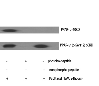
.jpg)
- 产品详情
- 实验流程
- 背景知识
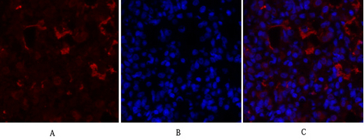
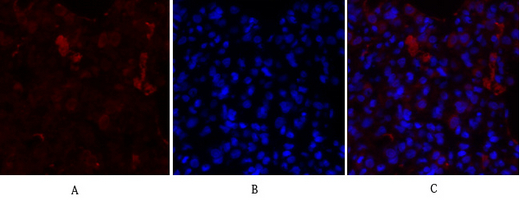
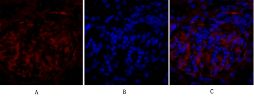
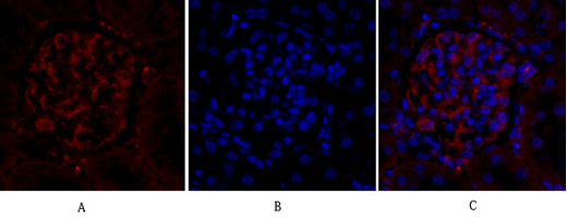
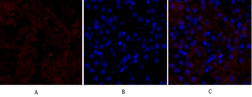
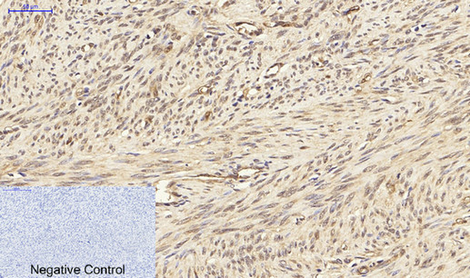
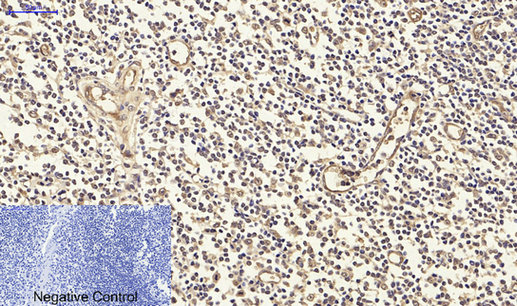
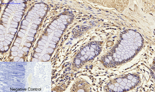
Application
| IF, ICC, WB, IHC-P, E |
|---|---|
| Primary Accession | P37231 |
| Reactivity | Human, Mouse, Rat |
| Host | Rabbit |
| Clonality | Polyclonal |
| Calculated MW | 57620 Da |
| Gene ID | 5468 |
|---|---|
| Other Names | PPARG; NR1C3; Peroxisome proliferator-activated receptor gamma; PPAR-gamma; Nuclear receptor subfamily 1 group C member 3 |
| Dilution | IF~~IF: 1:50-200 Western Blot: 1/500 - 1/2000. Immunohistochemistry: 1/100 - 1/300. ELISA: 1/10000. Not yet tested in other applications. ICC~~N/A WB~~IF: 1:50-200 Western Blot: 1/500 - 1/2000. Immunohistochemistry: 1/100 - 1/300. ELISA: 1/10000. Not yet tested in other applications. IHC-P~~IF: 1:50-200 Western Blot: 1/500 - 1/2000. Immunohistochemistry: 1/100 - 1/300. ELISA: 1/10000. Not yet tested in other applications. E~~N/A |
| Format | Liquid in PBS containing 50% glycerol, 0.5% BSA and 0.09% (W/V) sodium azide. |
| Storage Conditions | -20℃ |
| Name | PPARG |
|---|---|
| Synonyms | NR1C3 |
| Function | Ligand-activated transcription factor that forms obligate heterodimers with the retinoic acid receptor and acts as a key regulator of biological processes, such as adipocyte differentiation, lipid metabolism, glucose homeostasis and beta-oxidation of fatty acids (PubMed:16150867, PubMed:20829347, PubMed:23525231, PubMed:8702406, PubMed:8706692, PubMed:9065481). Activated by lipid ligands: binds peroxisome proliferators, such as hypolipidemic drugs, and fatty acids, such as prostaglandin J2 metabolites (PubMed:16150867, PubMed:20829347, PubMed:23525231, PubMed:8702406, PubMed:8706692, PubMed:9065481). Ligand-binding results in a conformational change in the receptor, promoting dissociation of repressors and recruitment of coactivators, and subsequent activation of target gene expression (PubMed:16150867, PubMed:20829347, PubMed:23525231, PubMed:8702406, PubMed:8706692, PubMed:9065481). Specifically binds to DNA specific PPAR response elements (PPRE) and modulates the transcription of its target genes, such as acyl-CoA oxidase (By similarity). Acts as a critical regulator of gut homeostasis by suppressing NF-kappa-B-mediated pro-inflammatory responses (PubMed:20829347). Plays a role in the regulation of cardiovascular circadian rhythms by regulating the transcription of BMAL1 in the blood vessels (By similarity). |
| Cellular Location | Nucleus. Cytoplasm Note=Redistributed from the nucleus to the cytosol through a MAP2K1/MEK1-dependent manner (PubMed:17101779). NOCT enhances its nuclear translocation (By similarity). {ECO:0000250|UniProtKB:P37238, ECO:0000269|PubMed:17101779} |
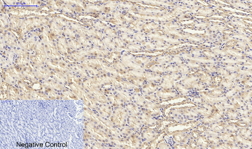
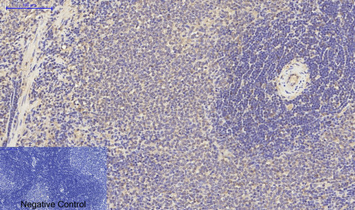
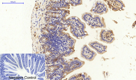
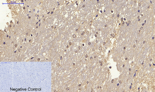
| Tissue Location | Highest expression in adipose tissue. Lower in skeletal muscle, spleen, heart and liver. Also detectable in placenta, lung and ovary. |
For Research Use Only. Not For Use In Diagnostic Procedures.
Provided below are standard protocols that you may find useful for product applications.
BACKGROUND
Nuclear receptor that binds peroxisome proliferators such as hypolipidemic drugs and fatty acids. Once activated by a ligand, the nuclear receptor binds to DNA specific PPAR response elements (PPRE) and modulates the transcription of its target genes, such as acyl-CoA oxidase. It therefore controls the peroxisomal beta-oxidation pathway of fatty acids. Key regulator of adipocyte differentiation and glucose homeostasis. ARF6 acts as a key regulator of the tissue-specific adipocyte P2 (aP2) enhancer. Acts as a critical regulator of gut homeostasis by suppressing NF-kappa-B-mediated proinflammatory responses. Plays a role in the regulation of cardiovascular circadian rhythms by regulating the transcription of ARNTL/BMAL1 in the blood vessels (By similarity).
终于等到您。ABCEPTA(百远生物)抗体产品。
点击下方“我要评价 ”按钮提交您的反馈信息,您的反馈和评价是我们最宝贵的财富之一,
我们将在1-3个工作日内处理您的反馈信息。
如有疑问,联系:0512-88856768 tech-china@abcepta.com.


