


 癌症的基本特征包括细胞增殖、血管生成、迁移、凋亡逃避机制和细胞永生等。找到癌症发生过程中这些通路的关键标记物和对应的抗体用于检测至关重要。
 为您推荐一个泛素化位点预测神器——泛素化分析工具,可以为您的蛋白的泛素化位点作出预测和评分。
 细胞自噬受体图形绘图工具为你的蛋白的细胞受体结合位点作出预测和评分,识别结合到自噬通路中的蛋白是非常重要的,便于让我们理解自噬在正常生理、病理过程中的作用,如发育、细胞分化、神经退化性疾病、压力条件下、感染和癌症。
MAPK10 Antibody (N-term)
Purified Rabbit Polyclonal Antibody (Pab)
- 产品详情
- 文献引用 : 1
- 实验流程
- 背景知识
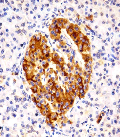
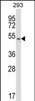
Application
| WB, IHC-P, E |
|---|---|
| Primary Accession | P53779 |
| Reactivity | Human, Rat, Mouse |
| Host | Rabbit |
| Clonality | Polyclonal |
| Isotype | Rabbit IgG |
| Calculated MW | 52585 Da |
| Antigen Region | 7-34 aa |
| Gene ID | 5602 |
|---|---|
| Other Names | Mitogen-activated protein kinase 10, MAP kinase 10, MAPK 10, MAP kinase p49 3F12, Stress-activated protein kinase 1b, SAPK1b, Stress-activated protein kinase JNK3, c-Jun N-terminal kinase 3, MAPK10, JNK3, JNK3A, PRKM10, SAPK1B |
| Target/Specificity | This MAPK10 antibody is generated from rabbits immunized with a KLH conjugated synthetic peptide between 7-34 amino acids from the N-terminal region of human MAPK10. |
| Dilution | WB~~1:1000 IHC-P~~1:100~500 E~~Use at an assay dependent concentration. |
| Format | Purified polyclonal antibody supplied in PBS with 0.09% (W/V) sodium azide. This antibody is purified through a protein A column, followed by peptide affinity purification. |
| Storage | Maintain refrigerated at 2-8°C for up to 2 weeks. For long term storage store at -20°C in small aliquots to prevent freeze-thaw cycles. |
| Precautions | MAPK10 Antibody (N-term) is for research use only and not for use in diagnostic or therapeutic procedures. |
| Name | MAPK10 |
|---|---|
| Synonyms | JNK3, JNK3A, PRKM10, SAPK1B |
| Function | Serine/threonine-protein kinase involved in various processes such as neuronal proliferation, differentiation, migration and programmed cell death. Extracellular stimuli such as pro-inflammatory cytokines or physical stress stimulate the stress-activated protein kinase/c-Jun N-terminal kinase (SAP/JNK) signaling pathway. In this cascade, two dual specificity kinases MAP2K4/MKK4 and MAP2K7/MKK7 phosphorylate and activate MAPK10/JNK3. In turn, MAPK10/JNK3 phosphorylates a number of transcription factors, primarily components of AP-1 such as JUN and ATF2 and thus regulates AP-1 transcriptional activity. Plays regulatory roles in the signaling pathways during neuronal apoptosis. Phosphorylates the neuronal microtubule regulator STMN2. Acts in the regulation of the amyloid-beta precursor protein/APP signaling during neuronal differentiation by phosphorylating APP. Also participates in neurite growth in spiral ganglion neurons. Phosphorylates the CLOCK-BMAL1 heterodimer and plays a role in the photic regulation of the circadian clock (PubMed:22441692). Phosphorylates JUND and this phosphorylation is inhibited in the presence of MEN1 (PubMed:22327296). |
| Cellular Location | Cytoplasm. Membrane; Lipid-anchor. Nucleus Mitochondrion. Note=Palmitoylation regulates MAPK10 trafficking to cytoskeleton. Recruited to the mitochondria in the presence of SARM1 (By similarity). |
| Tissue Location | Specific to a subset of neurons in the nervous system. Present in the hippocampus and areas, cerebellum, striatum, brain stem, and weakly in the spinal cord. Very weak expression in testis and kidney |
For Research Use Only. Not For Use In Diagnostic Procedures.

Provided below are standard protocols that you may find useful for product applications.
BACKGROUND
MAPK10 is a member of the MAP kinase family. MAP kinases act as an integration point for multiple biochemical signals, and are involved in a wide variety of cellular processes such as proliferation, differentiation, transcription regulation and development. This protein is a neuronal-specific form of c-Jun N-terminal kinases (JNKs). Through its phosphorylation and nuclear localization, this kinase plays regulatory roles in the signaling pathways during neuronal apoptosis. Beta-arrestin 2, a receptor-regulated MAP kinase scaffold protein, is found to interact with, and stimulate the phosphorylation of this kinase by MAP kinase kinase 4 (MKK4). Cyclin-dependent kinase 5 can phosphorylate, and inhibit the activity of this kinase, which may be important in preventing neuronal apoptosis.
REFERENCES
Li, B.S., et al., EMBO J. 21(3):324-333 (2002).
Yoshida, S., et al., J. Hum. Genet. 47(11):614-619 (2002).
McDonald, P.H., et al., Science 290(5496):1574-1577 (2000).
Yang, D.D., et al., Nature 389(6653):865-870 (1997).
Gupta, S., et al., EMBO J. 15(11):2760-2770 (1996).
终于等到您。ABCEPTA(百远生物)抗体产品。
点击下方“我要评价 ”按钮提交您的反馈信息,您的反馈和评价是我们最宝贵的财富之一,
我们将在1-3个工作日内处理您的反馈信息。
如有疑问,联系:0512-88856768 tech-china@abcepta.com.


