


 癌症的基本特征包括细胞增殖、血管生成、迁移、凋亡逃避机制和细胞永生等。找到癌症发生过程中这些通路的关键标记物和对应的抗体用于检测至关重要。
 为您推荐一个泛素化位点预测神器——泛素化分析工具,可以为您的蛋白的泛素化位点作出预测和评分。
 细胞自噬受体图形绘图工具为你的蛋白的细胞受体结合位点作出预测和评分,识别结合到自噬通路中的蛋白是非常重要的,便于让我们理解自噬在正常生理、病理过程中的作用,如发育、细胞分化、神经退化性疾病、压力条件下、感染和癌症。
CD148 Polyclonal Antibody
.jpg)
.jpg)
- 产品详情
- 实验流程
- 背景知识
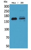
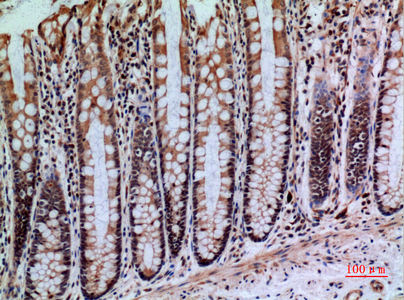
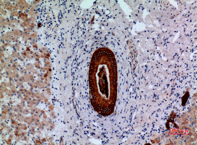
Application
| WB, IHC-P |
|---|---|
| Primary Accession | Q12913 |
| Reactivity | Human |
| Host | Rabbit |
| Clonality | Polyclonal |
| Calculated MW | 145941 Da |
| Gene ID | 5795 |
|---|---|
| Other Names | PTPRJ; DEP1; Receptor-type tyrosine-protein phosphatase eta; Protein-tyrosine phosphatase eta; R-PTP-eta; Density-enhanced phosphatase 1; DEP-1; HPTP eta; Protein-tyrosine phosphatase receptor type J; R-PTP-J; CD148 |
| Dilution | WB~~Western Blot: 1/500 - 1/2000. IHC-p: 1/100-1/300. ELISA: 1/20000. Not yet tested in other applications. IHC-P~~Western Blot: 1/500 - 1/2000. IHC-p: 1/100-1/300. ELISA: 1/20000. Not yet tested in other applications. |
| Format | Liquid in PBS containing 50% glycerol, 0.5% BSA and 0.09% (W/V) sodium azide. |
| Storage Conditions | -20℃ |
| Name | PTPRJ |
|---|---|
| Synonyms | DEP1 |
| Function | Tyrosine phosphatase which dephosphorylates or contributes to the dephosphorylation of CTNND1, FLT3, PDGFRB, MET, KDR, LYN, SRC, MAPK1, MAPK3, EGFR, TJP1, OCLN, PIK3R1 and PIK3R2 (PubMed:10821867, PubMed:12062403, PubMed:12370829, PubMed:12475979, PubMed:18348712, PubMed:19494114, PubMed:19922411, PubMed:21262971). Plays a role in cell adhesion, migration, proliferation and differentiation (PubMed:12370829, PubMed:14709717, PubMed:16682945, PubMed:19836242). Has a role in megakaryocytes and platelet formation (PubMed:30591527). Involved in vascular development (By similarity). Regulator of macrophage adhesion and spreading (By similarity). Positively affects cell-matrix adhesion (By similarity). Positive regulator of platelet activation and thrombosis. Negative regulator of cell proliferation (PubMed:16682945). Negative regulator of PDGF-stimulated cell migration; through dephosphorylation of PDGFR (PubMed:21091576). Positive regulator of endothelial cell survival, as well as of VEGF- induced SRC and AKT activation; through KDR dephosphorylation (PubMed:18936167). Negative regulator of EGFR signaling pathway; through EGFR dephosphorylation (PubMed:19836242). Enhances the barrier function of epithelial junctions during reassembly (PubMed:19332538). Negatively regulates T-cell receptor (TCR) signaling (PubMed:11259588, PubMed:9531590, PubMed:9780142). Upon T-cell TCR activation, it is up- regulated and excluded from the immunological synapses, while upon T- cell-antigen presenting cells (APC) disengagement, it is no longer excluded and can dephosphorylate PLCG1 and LAT to down-regulate prolongation of signaling (PubMed:11259588, PubMed:12913111). |
| Cellular Location | Cell membrane; Single-pass type I membrane protein. Cell projection, ruffle membrane. Cell junction Note=After T-cell stimulation, it is temporarily excluded from immunological synapses |
| Tissue Location | Expressed in the promyelocytic cell line HL-60, the granulocyte-macrophage colony-stimulating factor-dependent leukemic cell line F-36P, and the IL3 and erythropoietin-dependent leukemic cell line F-36E. Expressed predominantly in epithelial cells and lymphocytes. Enhanced expression at high cell density |
For Research Use Only. Not For Use In Diagnostic Procedures.
Provided below are standard protocols that you may find useful for product applications.
BACKGROUND
Tyrosine phosphatase which dephosphorylates or contributes to the dephosphorylation of CTNND1, FLT3, PDGFRB, MET, RET (variant MEN2A), KDR, LYN, SRC, MAPK1, MAPK3, EGFR, TJP1, OCLN, PIK3R1 and PIK3R2. Plays a role in cell adhesion, migration, proliferation and differentiation. Involved in vascular development. Regulator of macrophage adhesion and spreading. Positively affects cell-matrix adhesion. Positive regulator of platelet activation and thrombosis. Negative regulator of cell proliferation. Negative regulator of PDGF-stimulated cell migration; through dephosphorylation of PDGFR. Positive regulator of endothelial cell survival, as well as of VEGF-induced SRC and AKT activation; through KDR dephosphorylation. Negative regulator of EGFR signaling pathway; through EGFR dephosphorylation. Enhances the barrier function of epithelial junctions during reassembly. Negatively regulates T-cell receptor (TCR) signaling. Upon T-cell TCR activation, it is up-regulated and excluded from the immunological synapses, while upon T-cell-antigen presenting cells (APC) disengagement, it is no longer excluded and can dephosphorylate PLCG1 and LAT to down-regulate prolongation of signaling.
终于等到您。ABCEPTA(百远生物)抗体产品。
点击下方“我要评价 ”按钮提交您的反馈信息,您的反馈和评价是我们最宝贵的财富之一,
我们将在1-3个工作日内处理您的反馈信息。
如有疑问,联系:0512-88856768 tech-china@abcepta.com.


