


 癌症的基本特征包括细胞增殖、血管生成、迁移、凋亡逃避机制和细胞永生等。找到癌症发生过程中这些通路的关键标记物和对应的抗体用于检测至关重要。
 为您推荐一个泛素化位点预测神器——泛素化分析工具,可以为您的蛋白的泛素化位点作出预测和评分。
 细胞自噬受体图形绘图工具为你的蛋白的细胞受体结合位点作出预测和评分,识别结合到自噬通路中的蛋白是非常重要的,便于让我们理解自噬在正常生理、病理过程中的作用,如发育、细胞分化、神经退化性疾病、压力条件下、感染和癌症。
CD3-δ Polyclonal Antibody
.jpg)
- 产品详情
- 实验流程
- 背景知识
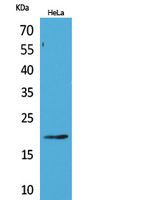
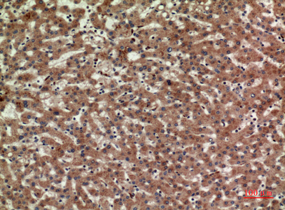
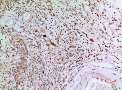
Application
| WB, IHC-P, IF, ICC, E |
|---|---|
| Primary Accession | P04234 |
| Reactivity | Human, Rat, Mouse |
| Host | Rabbit |
| Clonality | Polyclonal |
| Calculated MW | 18930 Da |
| Gene ID | 915 |
|---|---|
| Other Names | CD3D; T3D; T-cell surface glycoprotein CD3 delta chain; T-cell receptor T3 delta chain; CD3d |
| Dilution | WB~~Western Blot: 1/500 - 1/2000. IHC-p: 1/100-1/300. ELISA: 1/20000. Not yet tested in other applications. IHC-P~~Western Blot: 1/500 - 1/2000. IHC-p: 1/100-1/300. ELISA: 1/20000. Not yet tested in other applications. IF~~1:50~200 ICC~~N/A E~~N/A |
| Format | Liquid in PBS containing 50% glycerol, 0.5% BSA and 0.09% (W/V) sodium azide. |
| Storage Conditions | -20℃ |
| Name | CD3D |
|---|---|
| Synonyms | T3D |
| Function | Part of the TCR-CD3 complex present on T-lymphocyte cell surface that plays an essential role in adaptive immune response. When antigen presenting cells (APCs) activate T-cell receptor (TCR), TCR- mediated signals are transmitted across the cell membrane by the CD3 chains CD3D, CD3E, CD3G and CD247/CD3Z. All CD3 chains contain immunoreceptor tyrosine-based activation motifs (ITAMs) in their cytoplasmic domain. Upon TCR engagement, these motifs become phosphorylated by Src family protein tyrosine kinases LCK and FYN, resulting in the activation of downstream signaling pathways (PubMed:2470098). In addition of this role of signal transduction in T- cell activation, CD3D plays an essential role in thymocyte differentiation. Indeed, participates in correct intracellular TCR-CD3 complex assembly and surface expression. In absence of a functional TCR-CD3 complex, thymocytes are unable to differentiate properly. Interacts with CD4 and CD8 and thus serves to establish a functional link between the TCR and coreceptors CD4 and CD8, which is needed for activation and positive selection of CD4 or CD8 T-cells (PubMed:12215456). |
| Cellular Location | Cell membrane; Single-pass type I membrane protein |
| Tissue Location | CD3D is mostly present on T-lymphocytes with its TCR-CD3 partners. Present also in fetal NK-cells |
For Research Use Only. Not For Use In Diagnostic Procedures.
Provided below are standard protocols that you may find useful for product applications.
BACKGROUND
Part of the TCR-CD3 complex present on T-lymphocyte cell surface that plays an essential role in adaptive immune response. When antigen presenting cells (APCs) activate T-cell receptor (TCR), TCR-mediated signals are transmitted across the cell membrane by the CD3 chains CD3D, CD3E, CD3G and CD3Z. All CD3 chains contain immunoreceptor tyrosine-based activation motifs (ITAMs) in their cytoplasmic domain. Upon TCR engagement, these motifs become phosphorylated by Src family protein tyrosine kinases LCK and FYN, resulting in the activation of downstream signaling pathways (PubMed:2470098). In addition of this role of signal transduction in T-cell activation, CD3D plays an essential role in thymocyte differentiation. Indeed, participates in correct intracellular TCR-CD3 complex assembly and surface expression. In absence of a functional TCR-CD3 complex, thymocytes are unable to differentiate properly. Interacts with CD4 and CD8 and thus serves to establish a functional link between the TCR and coreceptors CD4 and CD8, which is needed for activation and positive selection of CD4 or CD8 T-cells(PubMed:12215456).
终于等到您。ABCEPTA(百远生物)抗体产品。
点击下方“我要评价 ”按钮提交您的反馈信息,您的反馈和评价是我们最宝贵的财富之一,
我们将在1-3个工作日内处理您的反馈信息。
如有疑问,联系:0512-88856768 tech-china@abcepta.com.


