


 癌症的基本特征包括细胞增殖、血管生成、迁移、凋亡逃避机制和细胞永生等。找到癌症发生过程中这些通路的关键标记物和对应的抗体用于检测至关重要。
 为您推荐一个泛素化位点预测神器——泛素化分析工具,可以为您的蛋白的泛素化位点作出预测和评分。
 细胞自噬受体图形绘图工具为你的蛋白的细胞受体结合位点作出预测和评分,识别结合到自噬通路中的蛋白是非常重要的,便于让我们理解自噬在正常生理、病理过程中的作用,如发育、细胞分化、神经退化性疾病、压力条件下、感染和癌症。
IL-1F8 Polyclonal Antibody
.jpg)
- 产品详情
- 实验流程
- 背景知识
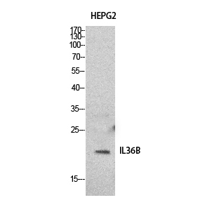
Application
| WB, IHC-P, IF, ICC, E |
|---|---|
| Primary Accession | Q9NZH7 |
| Reactivity | Human, Rat, Mouse |
| Host | Rabbit |
| Clonality | Polyclonal |
| Calculated MW | 18522 Da |
| Gene ID | 27177 |
|---|---|
| Other Names | IL36B; IL1F8; IL1H2; Interleukin-36 beta; FIL1 eta; Interleukin-1 eta; IL-1 eta; Interleukin-1 family member 8; IL-1F8; Interleukin-1 homolog 2; IL-1H2 |
| Dilution | WB~~Western Blot: 1/500 - 1/2000. IHC-p: 1/100-1/300. ELISA: 1/20000. Not yet tested in other applications. IHC-P~~Western Blot: 1/500 - 1/2000. IHC-p: 1/100-1/300. ELISA: 1/20000. Not yet tested in other applications. IF~~1:50~200 ICC~~N/A E~~N/A |
| Format | Liquid in PBS containing 50% glycerol, 0.5% BSA and 0.09% (W/V) sodium azide. |
| Storage Conditions | -20℃ |
| Name | IL36B (HGNC:15564) |
|---|---|
| Synonyms | IL1F8, IL1H2 |
| Function | Cytokine that binds to and signals through the IL1RL2/IL-36R receptor which in turn activates NF-kappa-B and MAPK signaling pathways in target cells linked to a pro-inflammatory response. Part of the IL- 36 signaling system that is thought to be present in epithelial barriers and to take part in local inflammatory response; similar to the IL-1 system with which it shares the coreceptor IL1RAP. Stimulates production of interleukin-6 and interleukin-8 in synovial fibrobasts, articular chondrocytes and mature adipocytes. Induces expression of a number of antimicrobial peptides including beta-defensins 4 and 103 as well as a number of matrix metalloproteases. Seems to be involved in skin inflammatory response by acting on keratinocytes, dendritic cells and indirectly on T-cells to drive tissue infiltration, cell maturation and cell proliferation. In cultured keratinocytes induces the expression of macrophage, T-cell, and neutrophil chemokines, such as CCL3, CCL4, CCL5, CCL2, CCL17, CCL22, CL20, CCL5, CCL2, CCL17, CCL22, CXCL8, CCL20 and CXCL1, and the production of pro-inflammatory cytokines such as TNF-alpha, IL-8 and IL-6. |
| Cellular Location | Cytoplasm. Secreted. Note=The secretion is dependent on protein unfolding and facilitated by the cargo receptor TMED10; it results in protein translocation from the cytoplasm into the ERGIC (endoplasmic reticulum-Golgi intermediate compartment) followed by vesicle entry and secretion. |
| Tissue Location | Expression at low levels in tonsil, bone marrow, heart, placenta, lung, testis and colon but not in any hematopoietic cell lines. Not detected in adipose tissue. Expressed at higher levels in psoriatic plaques than in symptomless psoriatic skin or healthy control skin. Increased levels are not detected in inflamed joint tissue. |
For Research Use Only. Not For Use In Diagnostic Procedures.
Provided below are standard protocols that you may find useful for product applications.
BACKGROUND
Cytokine that binds to and signals through the IL1RL2/IL-36R receptor which in turn activates NF-kappa-B and MAPK signaling pathways in target cells linked to a pro-inflammatory response. Part of the IL-36 signaling system that is thought to be present in epithelial barriers and to take part in local inflammatory response; similar to the IL-1 system with which it shares the coreceptor IL1RAP. Stimulates production of interleukin-6 and interleukin-8 in synovial fibrobasts, articular chondrocytes and mature adipocytes. Induces expression of a number of antimicrobial peptides including beta-defensins 4 and 103 as well as a number of matrix metalloproteases. Seems to be involved in skin inflammatory response by acting on keratinocytes, dendritic cells and indirectly on T-cells to drive tissue infiltration, cell maturation and cell proliferation. In cultured keratinocytes induces the expression of macrophage, T-cell, and neutrophil chemokines, such as CCL3, CCL4, CCL5, CCL2, CCL17, CCL22, CL20, CCL5, CCL2, CCL17, CCL22, CXCL8, CCL20 and CXCL1, and the production of proinflammatory cytokines such as TNF-alpha, IL- 8 and IL-6.
终于等到您。ABCEPTA(百远生物)抗体产品。
点击下方“我要评价 ”按钮提交您的反馈信息,您的反馈和评价是我们最宝贵的财富之一,
我们将在1-3个工作日内处理您的反馈信息。
如有疑问,联系:0512-88856768 tech-china@abcepta.com.


