


 癌症的基本特征包括细胞增殖、血管生成、迁移、凋亡逃避机制和细胞永生等。找到癌症发生过程中这些通路的关键标记物和对应的抗体用于检测至关重要。
 为您推荐一个泛素化位点预测神器——泛素化分析工具,可以为您的蛋白的泛素化位点作出预测和评分。
 细胞自噬受体图形绘图工具为你的蛋白的细胞受体结合位点作出预测和评分,识别结合到自噬通路中的蛋白是非常重要的,便于让我们理解自噬在正常生理、病理过程中的作用,如发育、细胞分化、神经退化性疾病、压力条件下、感染和癌症。
EHD3 Antibody (N-term)
Purified Rabbit Polyclonal Antibody (Pab)

- 产品详情
- 实验流程
- 背景知识
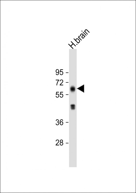
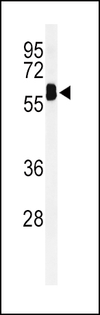
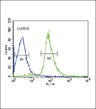
Application
| WB, IHC-P, FC, E |
|---|---|
| Primary Accession | Q9NZN3 |
| Other Accession | Q641Z6, Q9WVK4, Q9H4M9, Q5E9R3 |
| Reactivity | Human, Mouse |
| Predicted | Bovine, Mouse, Rat |
| Host | Rabbit |
| Clonality | Polyclonal |
| Isotype | Rabbit IgG |
| Calculated MW | 60887 Da |
| Antigen Region | 22-48 aa |
| Gene ID | 30845 |
|---|---|
| Other Names | EH domain-containing protein 3, PAST homolog 3, EHD3, EHD2, PAST3 |
| Target/Specificity | This EHD3 antibody is generated from rabbits immunized with a KLH conjugated synthetic peptide between 22-48 amino acids from the N-terminal region of human EHD3. |
| Dilution | WB~~1:1000 IHC-P~~1:100~500 FC~~1:10~50 E~~Use at an assay dependent concentration. |
| Format | Purified polyclonal antibody supplied in PBS with 0.09% (W/V) sodium azide. This antibody is prepared by Saturated Ammonium Sulfate (SAS) precipitation followed by dialysis against PBS. |
| Storage | Maintain refrigerated at 2-8°C for up to 2 weeks. For long term storage store at -20°C in small aliquots to prevent freeze-thaw cycles. |
| Precautions | EHD3 Antibody (N-term) is for research use only and not for use in diagnostic or therapeutic procedures. |
| Name | EHD3 (HGNC:3244) |
|---|---|
| Function | ATP- and membrane-binding protein that controls membrane reorganization/tubulation upon ATP hydrolysis (PubMed:25686250). In vitro causes tubulation of endocytic membranes (PubMed:24019528). Binding to phosphatidic acid induces its membrane tubulation activity (By similarity). Plays a role in endocytic transport. Involved in early endosome to recycling endosome compartment (ERC), retrograde early endosome to Golgi, and endosome to plasma membrane (rapid recycling) protein transport. Involved in the regulation of Golgi maintenance and morphology (PubMed:16251358, PubMed:17233914, PubMed:19139087, PubMed:23781025). Involved in the recycling of internalized D1 dopamine receptor (PubMed:21791287). Plays a role in cardiac protein trafficking probably implicating ANK2 (PubMed:20489164). Involved in the ventricular membrane targeting of SLC8A1 and CACNA1C and probably the atrial membrane localization of CACNA1GG and CACNA1H implicated in the regulation of atrial myocyte excitability and cardiac conduction (By similarity). In conjunction with EHD4 may be involved in endocytic trafficking of KDR/VEGFR2 implicated in control of glomerular function (By similarity). Involved in the rapid recycling of integrin beta-3 implicated in cell adhesion maintenance (PubMed:23781025). Involved in the unidirectional retrograde dendritic transport of endocytosed BACE1 and in efficient sorting of BACE1 to axons implicating a function in neuronal APP processing (By similarity). Plays a role in the formation of the ciliary vesicle, an early step in cilium biogenesis; possibly sharing redundant functions with EHD1 (PubMed:25686250). |
| Cellular Location | Recycling endosome membrane; Peripheral membrane protein; Cytoplasmic side. Cell membrane; Peripheral membrane protein; Cytoplasmic side. Cell projection, cilium membrane; Peripheral membrane protein; Cytoplasmic side. Note=Localizes to the ciliary pocket from where the cilium protrudes (PubMed:25686250) Colocalizes with RAB8A and MYO5B to a cytoplasmic tubular network devoid of RAB11A (By similarity). Colocalizes with ANK2 in myocyte perinuclear region (PubMed:20489164). Colocalizes with BACE1 in tubulovesicular cytoplasmic membranes. Colocalizes with BACE1 and APP amyloid beta proteins in hippocampal mossy fiber terminals (By similarity). {ECO:0000250|UniProtKB:Q9QXY6, ECO:0000269|PubMed:20489164, ECO:0000269|PubMed:25686250} |
| Tissue Location | Highly expressed in heart and brain and moderately expressed in kidney, liver, and placenta |
Research Areas
For Research Use Only. Not For Use In Diagnostic Procedures.
Application Protocols
Provided below are standard protocols that you may find useful for product applications.
BACKGROUND
EHD3 plays a role in endocytic transport.
REFERENCES
Soranzo, N., et al. Nat. Genet. 41(11):1182-1190(2009)
Naslavsky, N., et al. J. Cell. Sci. 122 (PT 3), 389-400 (2009)
Roland, J.T., et al. Mol. Biol. Cell 18(8):2828-2837(2007)
终于等到您。ABCEPTA(百远生物)抗体产品。
点击下方“我要评价 ”按钮提交您的反馈信息,您的反馈和评价是我们最宝贵的财富之一,
我们将在1-3个工作日内处理您的反馈信息。
如有疑问,联系:0512-88856768 tech-china@abcepta.com.
¥ 1,250.00
Cat# AP7458A


