


 癌症的基本特征包括细胞增殖、血管生成、迁移、凋亡逃避机制和细胞永生等。找到癌症发生过程中这些通路的关键标记物和对应的抗体用于检测至关重要。
 为您推荐一个泛素化位点预测神器——泛素化分析工具,可以为您的蛋白的泛素化位点作出预测和评分。
 细胞自噬受体图形绘图工具为你的蛋白的细胞受体结合位点作出预测和评分,识别结合到自噬通路中的蛋白是非常重要的,便于让我们理解自噬在正常生理、病理过程中的作用,如发育、细胞分化、神经退化性疾病、压力条件下、感染和癌症。
Polyclonal Antibody to Interleukin 1 Alpha (IL1a)
Interleukin 1 Alpha
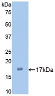
- 产品详情
- 实验流程
Application 
| WB |
|---|---|
| Primary Accession | P04822 |
| Reactivity | Rabbit |
| Host | Guinea Pig |
| Clonality | Polyclonal |
| Calculated MW | 30375 Da |
| Gene ID | 100009250 |
|---|---|
| Other Names | IL1-A; IL-1α; IL1F1; Preinterleukin 1 Alpha; Hematopoietin-1; Pro-Interleukin-1-Alpha; Interleukin-1 Family Member 1 |
| Target/Specificity | Oryctolagus cuniculus (Rabbit) |
| Dilution | WB~~Western blotting: 0.5-2ug/ml Immunocytochemistry in formalin fixed cells: 5-20ug/ml Immunohistochemistry in formalin fixed frozen section: 5-20ug/ml Immunohistochemistry in paraffin section: 5-20ug/ml Enzyme-linked Immunosorbent Assay: 0.05-2ug/ml Optimal working dilutions must be determined by end user. |
| Format | PBS, pH7.4, containing 0.02% NaN3, 50% glycerol. |
| Storage | Store at -20 °C.Stable for 12 months from date of receipt |
For Research Use Only. Not For Use In Diagnostic Procedures.
| Name | IL1A |
|---|---|
| Function | Cytokine constitutively present intracellularly in nearly all resting non-hematopoietic cells that plays an important role in inflammation and bridges the innate and adaptive immune systems. After binding to its receptor IL1R1 together with its accessory protein IL1RAP, forms the high affinity interleukin-1 receptor complex. Signaling involves the recruitment of adapter molecules such as MYD88, IRAK1 or IRAK4. In turn, mediates the activation of NF-kappa-B and the three MAPK pathways p38, p42/p44 and JNK pathways. Within the cell, acts as an alarmin and cell death results in its liberation in the extracellular space after disruption of the cell membrane to induce inflammation and alert the host to injury or damage. In addition to its role as a danger signal, which occurs when the cytokine is passively released by cell necrosis, directly senses DNA damage and acts as signal for genotoxic stress without loss of cell integrity. |
| Cellular Location | Nucleus {ECO:0000250|UniProtKB:P01583}. Cytoplasm {ECO:0000250|UniProtKB:P01583}. Secreted {ECO:0000250|UniProtKB:P01583}. Note=The lack of a specific hydrophobic segment in the precursor sequence suggests that IL-1 is released by damaged cells or is secreted by a mechanism differing from that used for other secretory proteins. The secretion is dependent on protein unfolding and facilitated by the cargo receptor TMED10; it results in protein translocation from the cytoplasm into the ERGIC (endoplasmic reticulum-Golgi intermediate compartment) followed by vesicle entry and secretion. Recruited to DNA damage sites and secreted after genotoxic stress. {ECO:0000250|UniProtKB:P01583} |
Research Areas
Application Protocols
Provided below are standard protocols that you may find useful for product applications.

终于等到您。ABCEPTA(百远生物)抗体产品。
点击下方“我要评价 ”按钮提交您的反馈信息,您的反馈和评价是我们最宝贵的财富之一,
我们将在1-3个工作日内处理您的反馈信息。
如有疑问,联系:0512-88856768 tech-china@abcepta.com.


