


 癌症的基本特征包括细胞增殖、血管生成、迁移、凋亡逃避机制和细胞永生等。找到癌症发生过程中这些通路的关键标记物和对应的抗体用于检测至关重要。
 为您推荐一个泛素化位点预测神器——泛素化分析工具,可以为您的蛋白的泛素化位点作出预测和评分。
 细胞自噬受体图形绘图工具为你的蛋白的细胞受体结合位点作出预测和评分,识别结合到自噬通路中的蛋白是非常重要的,便于让我们理解自噬在正常生理、病理过程中的作用,如发育、细胞分化、神经退化性疾病、压力条件下、感染和癌症。
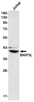
BNIP3L Rabbit mAb
- 产品详情
- 实验流程
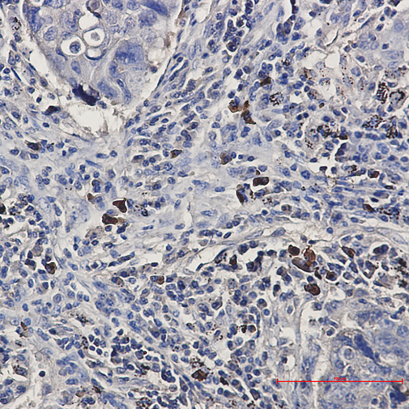
Application
| WB, IHC-P |
|---|---|
| Primary Accession | O60238 |
| Reactivity | Human |
| Host | Rabbit |
| Clonality | Monoclonal Antibody |
| Calculated MW | 23930 Da |
| Gene ID | 665 |
|---|---|
| Other Names | BNIP3L |
| Dilution | WB~~1/500-1/1000 IHC-P~~1:50~200 |
| Format | 50mM Tris-Glycine(pH 7.4), 0.15M NaCl, 40%Glycerol, 0.01% sodium azide and 0.05% BSA. |
| Storage | Store at 4°C short term. Aliquot and store at -20°C long term. Avoid freeze/thaw cycles. |
| Name | BNIP3L |
|---|---|
| Synonyms | BNIP3A, BNIP3H, NIX |
| Function | Induces apoptosis. Interacts with viral and cellular anti- apoptosis proteins. Can overcome the suppressors BCL-2 and BCL-XL, although high levels of BCL-XL expression will inhibit apoptosis. Inhibits apoptosis induced by BNIP3. Involved in mitochondrial quality control via its interaction with SPATA18/MIEAP: in response to mitochondrial damage, participates in mitochondrial protein catabolic process (also named MALM) leading to the degradation of damaged proteins inside mitochondria. The physical interaction of SPATA18/MIEAP, BNIP3 and BNIP3L/NIX at the mitochondrial outer membrane regulates the opening of a pore in the mitochondrial double membrane in order to mediate the translocation of lysosomal proteins from the cytoplasm to the mitochondrial matrix. May function as a tumor suppressor. |
| Cellular Location | Nucleus envelope. Endoplasmic reticulum. Mitochondrion outer membrane. Membrane; Single-pass membrane protein. Note=Colocalizes with SPATA18 at the mitochondrion outer membrane |
Research Areas
For Research Use Only. Not For Use In Diagnostic Procedures.
Application Protocols
Provided below are standard protocols that you may find useful for product applications.
终于等到您。ABCEPTA(百远生物)抗体产品。
点击下方“我要评价 ”按钮提交您的反馈信息,您的反馈和评价是我们最宝贵的财富之一,
我们将在1-3个工作日内处理您的反馈信息。
如有疑问,联系:0512-88856768 tech-china@abcepta.com.
¥ 1,500.00
Cat# AP75162


