


 癌症的基本特征包括细胞增殖、血管生成、迁移、凋亡逃避机制和细胞永生等。找到癌症发生过程中这些通路的关键标记物和对应的抗体用于检测至关重要。
 为您推荐一个泛素化位点预测神器——泛素化分析工具,可以为您的蛋白的泛素化位点作出预测和评分。
 细胞自噬受体图形绘图工具为你的蛋白的细胞受体结合位点作出预测和评分,识别结合到自噬通路中的蛋白是非常重要的,便于让我们理解自噬在正常生理、病理过程中的作用,如发育、细胞分化、神经退化性疾病、压力条件下、感染和癌症。
HLA C Rabbit mAb
- 产品详情
- 实验流程
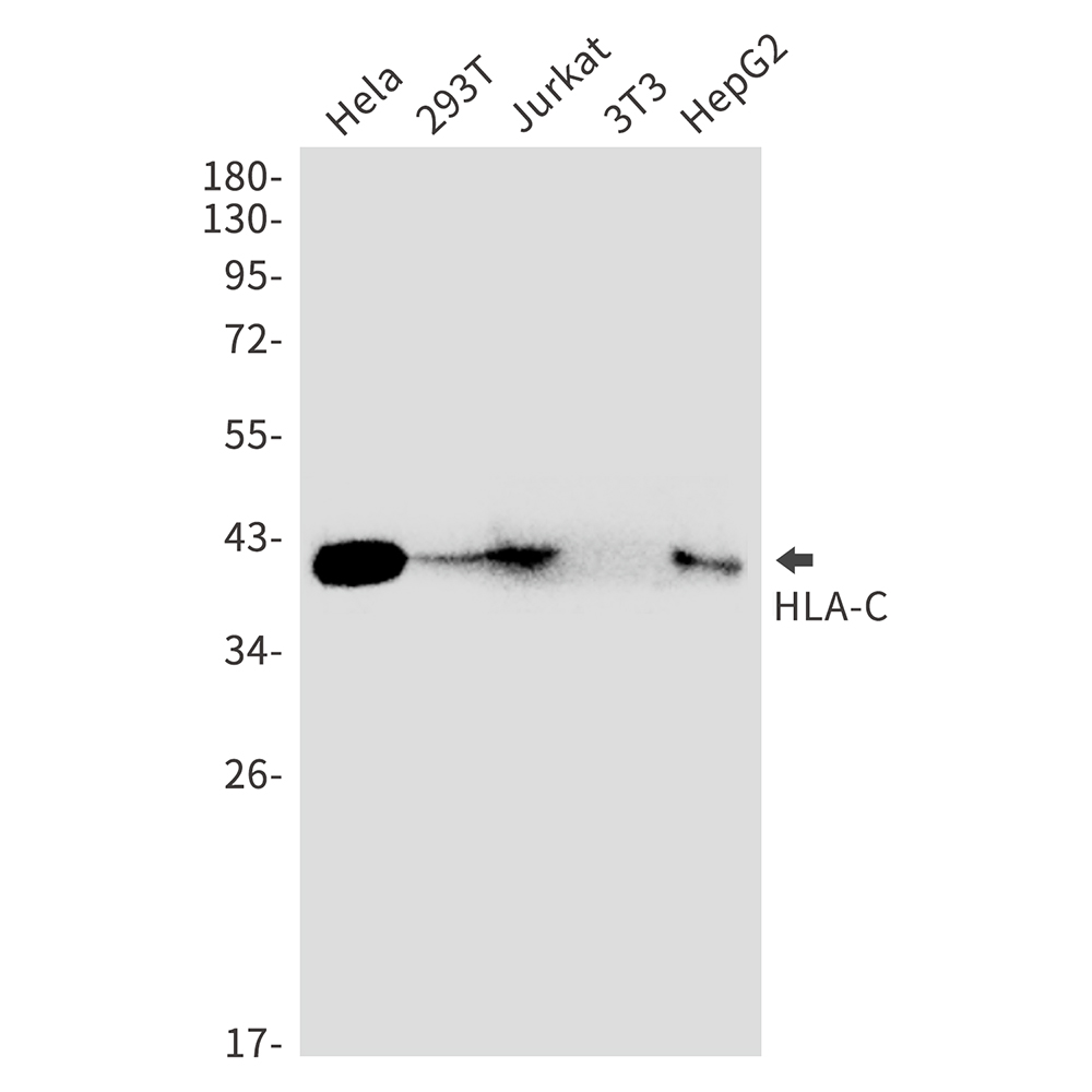
Application
| WB, IP |
|---|---|
| Primary Accession | P10321 |
| Reactivity | Human, Mouse |
| Host | Rabbit |
| Clonality | Monoclonal Antibody |
| Calculated MW | 40649 Da |
| Gene ID | 3107 |
|---|---|
| Other Names | HLA-C |
| Dilution | WB~~1/500-1/1000 IP~~N/A |
| Format | 50mM Tris-Glycine(pH 7.4), 0.15M NaCl, 40%Glycerol, 0.01% sodium azide and 0.05% BSA. |
| Storage | Store at 4°C short term. Aliquot and store at -20°C long term. Avoid freeze/thaw cycles. |
| Name | HLA-C (HGNC:4933) |
|---|---|
| Synonyms | HLAC |
| Function | Antigen-presenting major histocompatibility complex class I (MHCI) molecule with an important role in reproduction and antiviral immunity (PubMed:11172028, PubMed:20104487, PubMed:20439706, PubMed:20972337, PubMed:24091323, PubMed:28649982, PubMed:29312307). In complex with B2M/beta 2 microglobulin displays a restricted repertoire of self and viral peptides and acts as a dominant ligand for inhibitory and activating killer immunoglobulin receptors (KIRs) expressed on NK cells (PubMed:16141329). In an allogeneic setting, such as during pregnancy, mediates interaction of extravillous trophoblasts with KIR on uterine NK cells and regulate trophoblast invasion necessary for placentation and overall fetal growth (PubMed:20972337, PubMed:24091323). During viral infection, may present viral peptides with low affinity for KIRs, impeding KIR-mediated inhibition through peptide antagonism and favoring lysis of infected cells (PubMed:20439706). Presents a restricted repertoire of viral peptides on antigen-presenting cells for recognition by alpha-beta T cell receptor (TCR) on HLA-C-restricted CD8-positive T cells, guiding antigen-specific T cell immune response to eliminate infected cells, particularly in chronic viral infection settings such as HIV-1 or CMV infection (PubMed:11172028, PubMed:20104487, PubMed:28649982). Both the peptide and the MHC molecule are recognized by TCR, the peptide is responsible for the fine specificity of antigen recognition and MHC residues account for the MHC restriction of T cells (By similarity). Typically presents intracellular peptide antigens of 9 amino acids that arise from cytosolic proteolysis via proteasome. Can bind different peptides containing allele-specific binding motifs, which are mainly defined by anchor residues at position 2 and 9. Preferentially displays peptides having a restricted repertoire of hydrophobic or aromatic amino acids (Phe, Ile, Leu, Met, Val and Tyr) at the C-terminal anchor (PubMed:25311805, PubMed:8265661). |
| Cellular Location | Cell membrane; Single-pass type I membrane protein. Endoplasmic reticulum membrane; Single-pass membrane protein |
| Tissue Location | Ubiquitous. Highly expressed in fetal extravillous trophoblasts in the decidua basalis (at protein level) |
Research Areas
For Research Use Only. Not For Use In Diagnostic Procedures.
Application Protocols
Provided below are standard protocols that you may find useful for product applications.
终于等到您。ABCEPTA(百远生物)抗体产品。
点击下方“我要评价 ”按钮提交您的反馈信息,您的反馈和评价是我们最宝贵的财富之一,
我们将在1-3个工作日内处理您的反馈信息。
如有疑问,联系:0512-88856768 tech-china@abcepta.com.
¥ 1,500.00
Cat# AP75551


