


 癌症的基本特征包括细胞增殖、血管生成、迁移、凋亡逃避机制和细胞永生等。找到癌症发生过程中这些通路的关键标记物和对应的抗体用于检测至关重要。
 为您推荐一个泛素化位点预测神器——泛素化分析工具,可以为您的蛋白的泛素化位点作出预测和评分。
 细胞自噬受体图形绘图工具为你的蛋白的细胞受体结合位点作出预测和评分,识别结合到自噬通路中的蛋白是非常重要的,便于让我们理解自噬在正常生理、病理过程中的作用,如发育、细胞分化、神经退化性疾病、压力条件下、感染和癌症。
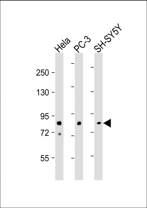
DYRK1A Antibody (N-term)
Purified Rabbit Polyclonal Antibody (Pab)
- 产品详情
- 实验流程
- 背景知识
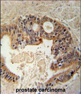
Application
| IHC-P, WB, E |
|---|---|
| Primary Accession | Q13627 |
| Other Accession | Q2TAE3, Q63470, Q61214 |
| Reactivity | Human, Mouse |
| Predicted | Mouse, Rat, Xenopus |
| Host | Rabbit |
| Clonality | Polyclonal |
| Isotype | Rabbit IgG |
| Calculated MW | 85584 Da |
| Antigen Region | 107-136 aa |
| Gene ID | 1859 |
|---|---|
| Other Names | Dual specificity tyrosine-phosphorylation-regulated kinase 1A, Dual specificity YAK1-related kinase, HP86, Protein kinase minibrain homolog, MNBH, hMNB, DYRK1A, DYRK, MNB, MNBH |
| Target/Specificity | This DYRK1A antibody is generated from rabbits immunized with a KLH conjugated synthetic peptide between 107-136 amino acids from the N-terminal region of human DYRK1A. |
| Dilution | IHC-P~~1:100~500 WB~~1:500-1:2000 E~~Use at an assay dependent concentration. |
| Format | Purified polyclonal antibody supplied in PBS with 0.09% (W/V) sodium azide. This antibody is prepared by Saturated Ammonium Sulfate (SAS) precipitation followed by dialysis against PBS. |
| Storage | Maintain refrigerated at 2-8°C for up to 2 weeks. For long term storage store at -20°C in small aliquots to prevent freeze-thaw cycles. |
| Precautions | DYRK1A Antibody (N-term) is for research use only and not for use in diagnostic or therapeutic procedures. |
| Name | DYRK1A {ECO:0000303|PubMed:25620562, ECO:0000312|HGNC:HGNC:3091} |
|---|---|
| Function | Dual-specificity kinase which possesses both serine/threonine and tyrosine kinase activities (PubMed:20981014, PubMed:21127067, PubMed:23665168, PubMed:30773093, PubMed:8769099). Exhibits a substrate preference for proline at position P+1 and arginine at position P-3 (PubMed:23665168). Plays an important role in double-strand breaks (DSBs) repair following DNA damage (PubMed:31024071). Mechanistically, phosphorylates RNF169 and increases its ability to block accumulation of TP53BP1 at the DSB sites thereby promoting homologous recombination repair (HRR) (PubMed:30773093). Also acts as a positive regulator of transcription by acting as a CTD kinase that mediates phosphorylation of the CTD (C-terminal domain) of the large subunit of RNA polymerase II (RNAP II) POLR2A (PubMed:25620562, PubMed:29849146). May play a role in a signaling pathway regulating nuclear functions of cell proliferation (PubMed:14500717). Modulates alternative splicing by phosphorylating the splice factor SRSF6 (By similarity). Has pro- survival function and negatively regulates the apoptotic process (By similarity). Promotes cell survival upon genotoxic stress through phosphorylation of SIRT1 (By similarity). This in turn inhibits p53/TP53 activity and apoptosis (By similarity). Phosphorylates SEPTIN4, SEPTIN5 and SF3B1 at 'Thr-434' (By similarity). |
| Cellular Location | Nucleus. Nucleus speckle {ECO:0000250|UniProtKB:Q61214} |
| Tissue Location | Ubiquitous. Highest levels in skeletal muscle, testis, fetal lung and fetal kidney. |
For Research Use Only. Not For Use In Diagnostic Procedures.
Provided below are standard protocols that you may find useful for product applications.
BACKGROUND
DYRK1A is a member of the Dual-specificity tyrosine phosphorylation-regulated kinase (DYRK) family. This member contains a nuclear targeting signal sequence, a protein kinase domain, a leucine zipper motif, and a highly conservative 13-consecutive-histidine repeat. It catalyzes its autophosphorylation on serine/threonine and tyrosine residues. It may play a significant role in a signaling pathway regulating cell proliferation and may be involved in brain development. The DYRK1A gene is a homolog of Drosophila mnb (minibrain) gene and rat Dyrk gene. It is localized in the Down syndrome critical region of chromosome 21, and is considered to be a strong candidate gene for learning defects associated with Down syndrome.
REFERENCES
Adayev,T., Biochemistry 46 (25), 7614-7624 (2007)
Chang,H.S., Int. J. Cancer 120 (11), 2377-2385 (2007)
Alvarez,M., Mol. Biol. Cell 18 (4), 1167-1178 (2007)
Wissing,J., Mol. Cell Proteomics 6 (3), 537-547 (2007)
终于等到您。ABCEPTA(百远生物)抗体产品。
点击下方“我要评价 ”按钮提交您的反馈信息,您的反馈和评价是我们最宝贵的财富之一,
我们将在1-3个工作日内处理您的反馈信息。
如有疑问,联系:0512-88856768 tech-china@abcepta.com.


