


 癌症的基本特征包括细胞增殖、血管生成、迁移、凋亡逃避机制和细胞永生等。找到癌症发生过程中这些通路的关键标记物和对应的抗体用于检测至关重要。
 为您推荐一个泛素化位点预测神器——泛素化分析工具,可以为您的蛋白的泛素化位点作出预测和评分。
 细胞自噬受体图形绘图工具为你的蛋白的细胞受体结合位点作出预测和评分,识别结合到自噬通路中的蛋白是非常重要的,便于让我们理解自噬在正常生理、病理过程中的作用,如发育、细胞分化、神经退化性疾病、压力条件下、感染和癌症。
MLKL Rabbit mAb
- 产品详情
- 实验流程
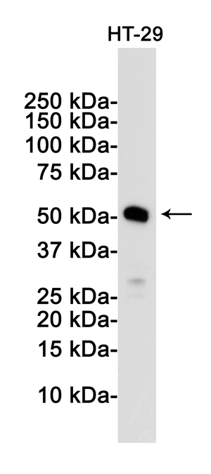
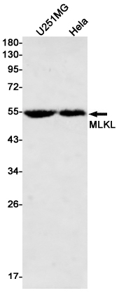
Application
| WB |
|---|---|
| Primary Accession | Q8NB16 |
| Reactivity | Human |
| Host | Rabbit |
| Clonality | Monoclonal Antibody |
| Calculated MW | 54479 Da |
| Gene ID | 197259 |
|---|---|
| Other Names | MLKL |
| Dilution | WB~~1/500-1/1000 |
| Format | 50mM Tris-Glycine(pH 7.4), 0.15M NaCl, 40%Glycerol, 0.01% sodium azide and 0.05% BSA. |
| Storage | Store at 4°C short term. Aliquot and store at -20°C long term. Avoid freeze/thaw cycles. |
| Name | MLKL {ECO:0000303|PubMed:22265413, ECO:0000312|HGNC:HGNC:26617} |
|---|---|
| Function | Pseudokinase that plays a key role in TNF-induced necroptosis, a programmed cell death process (PubMed:22265413, PubMed:22265414, PubMed:22421439, PubMed:24316671). Does not have protein kinase activity (PubMed:22265413, PubMed:22265414, PubMed:22421439, PubMed:24316671). Activated following phosphorylation by RIPK3, leading to homotrimerization, localization to the plasma membrane and execution of programmed necrosis characterized by calcium influx and plasma membrane damage (PubMed:22265413, PubMed:22265414, PubMed:22421439, PubMed:24316671). In addition to TNF-induced necroptosis, necroptosis can also take place in the nucleus in response to orthomyxoviruses infection: following activation by ZBP1, MLKL is phosphorylated by RIPK3 in the nucleus, triggering disruption of the nuclear envelope and leakage of cellular DNA into the cytosol.following ZBP1 activation, which senses double-stranded Z-RNA structures, nuclear RIPK3 catalyzes phosphorylation and activation of MLKL, promoting disruption of the nuclear envelope and leakage of cellular DNA into the cytosol (By similarity). Binds to highly phosphorylated inositol phosphates such as inositolhexakisphosphate (InsP6) which is essential for its necroptotic function (PubMed:29883610). |
| Cellular Location | Cytoplasm. Cell membrane Nucleus {ECO:0000250|UniProtKB:Q9D2Y4}. Note=Localizes to the cytoplasm and translocates to the plasma membrane on necroptosis induction (PubMed:24316671). Localizes to the nucleus in response to orthomyxoviruses infection (By similarity) {ECO:0000250|UniProtKB:Q9D2Y4, ECO:0000269|PubMed:24316671} |
Research Areas
For Research Use Only. Not For Use In Diagnostic Procedures.
Application Protocols
Provided below are standard protocols that you may find useful for product applications.
终于等到您。ABCEPTA(百远生物)抗体产品。
点击下方“我要评价 ”按钮提交您的反馈信息,您的反馈和评价是我们最宝贵的财富之一,
我们将在1-3个工作日内处理您的反馈信息。
如有疑问,联系:0512-88856768 tech-china@abcepta.com.
¥ 1,500.00
Cat# AP75727


