


 癌症的基本特征包括细胞增殖、血管生成、迁移、凋亡逃避机制和细胞永生等。找到癌症发生过程中这些通路的关键标记物和对应的抗体用于检测至关重要。
 为您推荐一个泛素化位点预测神器——泛素化分析工具,可以为您的蛋白的泛素化位点作出预测和评分。
 细胞自噬受体图形绘图工具为你的蛋白的细胞受体结合位点作出预测和评分,识别结合到自噬通路中的蛋白是非常重要的,便于让我们理解自噬在正常生理、病理过程中的作用,如发育、细胞分化、神经退化性疾病、压力条件下、感染和癌症。
CABP1 Antibody (C-term)
Purified Rabbit Polyclonal Antibody (Pab)
- 产品详情
- 实验流程
- 背景知识
Application
| WB, IHC-P, E |
|---|---|
| Primary Accession | Q9NZU7 |
| Other Accession | O88751, Q9JLK7, Q9N1R0 |
| Reactivity | Human, Mouse, Rat |
| Predicted | Rat, Bovine |
| Host | Rabbit |
| Clonality | Polyclonal |
| Isotype | Rabbit IgG |
| Calculated MW | 39838 Da |
| Antigen Region | 311-343 aa |
| Gene ID | 9478 |
|---|---|
| Other Names | Calcium-binding protein 1, CaBP1, Calbrain, Caldendrin, CABP1 |
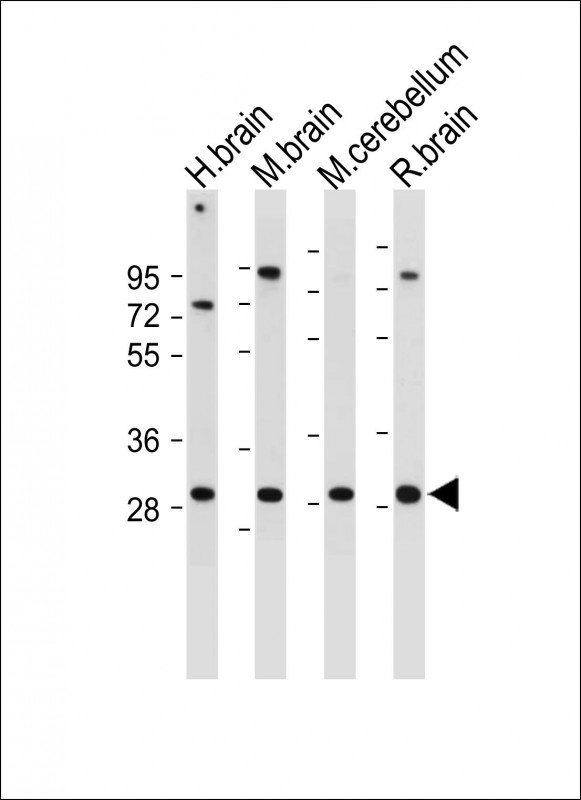
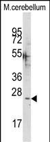
| Target/Specificity | This CABP1 antibody is generated from rabbits immunized with a KLH conjugated synthetic peptide between 311-343 amino acids from the C-terminal region of human CABP1. |
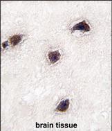
| Dilution | WB~~1:1000 IHC-P~~1:100~500 E~~Use at an assay dependent concentration. |
| Format | Purified polyclonal antibody supplied in PBS with 0.09% (W/V) sodium azide. This antibody is purified through a protein A column, followed by peptide affinity purification. |
| Storage | Maintain refrigerated at 2-8°C for up to 2 weeks. For long term storage store at -20°C in small aliquots to prevent freeze-thaw cycles. |
| Precautions | CABP1 Antibody (C-term) is for research use only and not for use in diagnostic or therapeutic procedures. |
| Name | CABP1 |
|---|---|
| Function | Modulates calcium-dependent activity of inositol 1,4,5- triphosphate receptors (ITPRs) (PubMed:14570872). Inhibits agonist- induced intracellular calcium signaling (PubMed:15980432). Enhances inactivation and does not support calcium-dependent facilitation of voltage-dependent P/Q-type calcium channels (PubMed:11865310). Causes calcium-dependent facilitation and inhibits inactivation of L-type calcium channels by binding to the same sites as calmodulin in the C- terminal domain of CACNA1C, but has an opposite effect on channel function (PubMed:15140941). Suppresses the calcium-dependent inactivation of CACNA1D (By similarity). Inhibits TRPC5 channels (PubMed:15895247). Prevents NMDA receptor-induced cellular degeneration. Required for the normal transfer of light signals through the retina (By similarity). |
| Cellular Location | Cytoplasm, cytoskeleton. Cytoplasm, perinuclear region. Cell membrane; Lipid-anchor; Cytoplasmic side. Golgi apparatus Postsynaptic density. Note=L-CaBP1 is associated most likely with the cytoskeletal structures, whereas S-CaBP1 is localized at or near the plasma membrane. [Isoform S-CaBP1]: Cytoplasm, cell cortex. Cell membrane; Lipid-anchor Note=S-CaBP1 is localized at or near the plasma membrane |
| Tissue Location | Retina and brain. Somatodendritic compartment of neurons. Calbrain was found exclusively in brain where it is abundant in the hippocampus, habenular area in the epithalamus and in the cerebellum |
For Research Use Only. Not For Use In Diagnostic Procedures.
Provided below are standard protocols that you may find useful for product applications.
BACKGROUND
CABP1 belongs to a subfamily of calcium binding proteins, which share similarity to calmodulin. Calcium binding proteins are an important component of calcium mediated cellular signal transduction. Expression of this protein was only detected in retina and brain. Study of the mouse homolog demonstrated that groups of cells expressing this protein are located in the center or inner border of the inner unclear layer of retina.
REFERENCES
Haynes,L.P., Proteomics 6 (6), 1822-1832 (2006) Wingard,J.N., J. Biol. Chem. 280 (45), 37461-37470 (2005) Zhou,H., J. Biol. Chem. 280 (33), 29612-29619 (2005) Haeseleer,F., J. Biol. Chem. 275 (2), 1247-1260 (2000)
终于等到您。ABCEPTA(百远生物)抗体产品。
点击下方“我要评价 ”按钮提交您的反馈信息,您的反馈和评价是我们最宝贵的财富之一,
我们将在1-3个工作日内处理您的反馈信息。
如有疑问,联系:0512-88856768 tech-china@abcepta.com.


