


 癌症的基本特征包括细胞增殖、血管生成、迁移、凋亡逃避机制和细胞永生等。找到癌症发生过程中这些通路的关键标记物和对应的抗体用于检测至关重要。
 为您推荐一个泛素化位点预测神器——泛素化分析工具,可以为您的蛋白的泛素化位点作出预测和评分。
 细胞自噬受体图形绘图工具为你的蛋白的细胞受体结合位点作出预测和评分,识别结合到自噬通路中的蛋白是非常重要的,便于让我们理解自噬在正常生理、病理过程中的作用,如发育、细胞分化、神经退化性疾病、压力条件下、感染和癌症。
Prostaglandin D Synthase Rabbit mAb
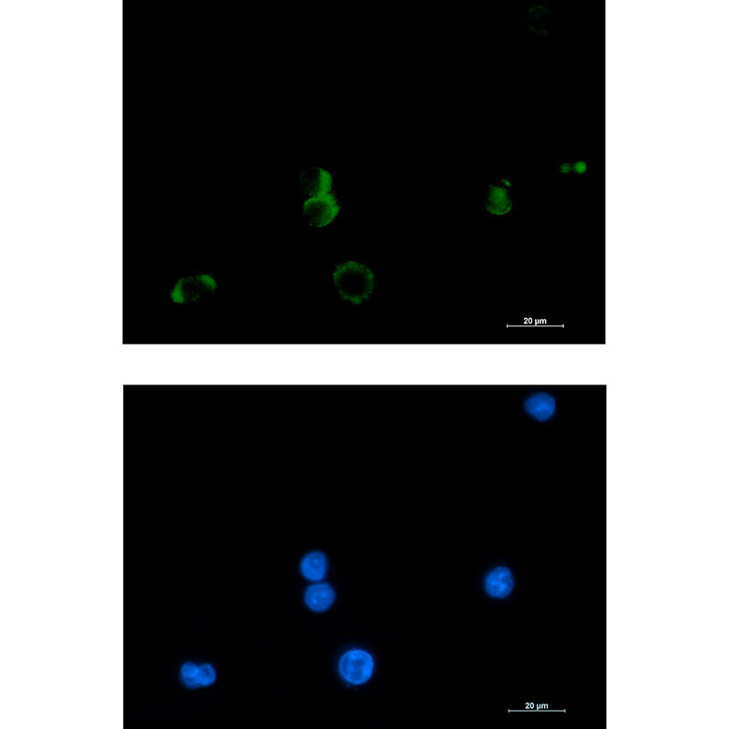
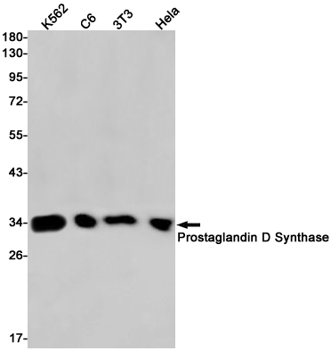
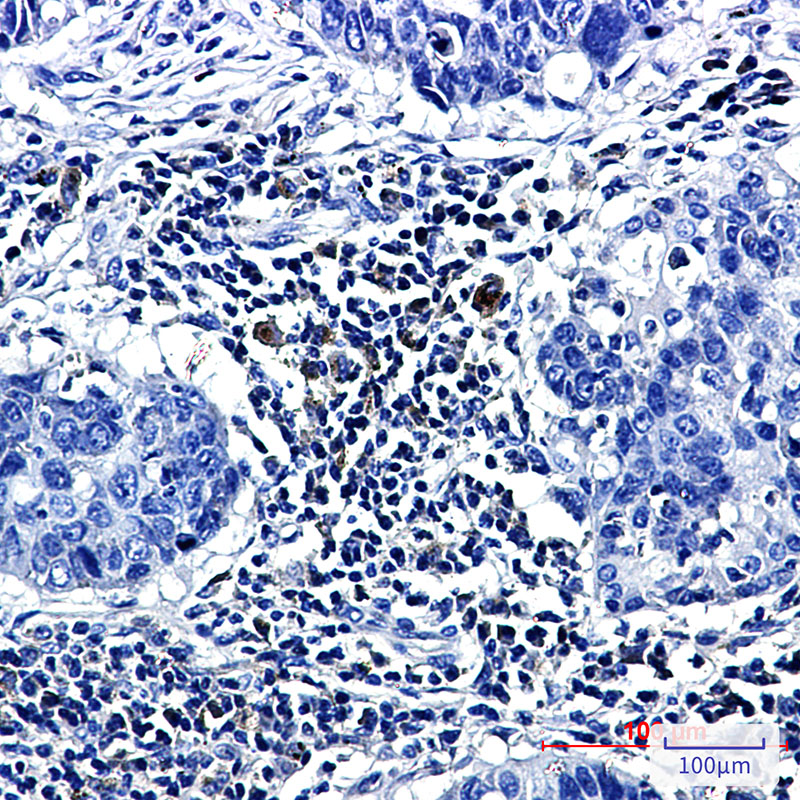
- 产品详情
- 实验流程
- 背景知识
Application 
| WB, IHC-P, IHC-F |
|---|---|
| Primary Accession | P41222 |
| Reactivity | Human |
| Host | Rabbit |
| Clonality | Monoclonal Antibody |
| Isotype | IgG |
| Conjugate | Unconjugated |
| Purification | Affinity Purified |
| Calculated MW | 21029 Da |
| Gene ID | 5730 |
|---|---|
| Other Names | PTGDS |
| Dilution | WB~~1:1000-1:5000 IHC-P~~1:50~200 IHC-F~~N/A |
| Format | Liquid in 50mM Tris-Glycine(pH 7.4), 0.15M NaCl, 40%Glycerol, 0.01% sodium azide and 0.05% BSA. |
| Storage | Store at 4°C short term. Aliquot and store at -20°C long term. Avoid freeze/thaw cycles. |
For Research Use Only. Not For Use In Diagnostic Procedures.
| Name | PTGDS |
|---|---|
| Synonyms | PDS |
| Function | Catalyzes the conversion of PGH2 to PGD2, a prostaglandin involved in smooth muscle contraction/relaxation and a potent inhibitor of platelet aggregation (PubMed:20667974). Involved in a variety of CNS functions, such as sedation, NREM sleep and PGE2-induced allodynia, and may have an anti-apoptotic role in oligodendrocytes. Binds small non- substrate lipophilic molecules, including biliverdin, bilirubin, retinal, retinoic acid and thyroid hormone, and may act as a scavenger for harmful hydrophobic molecules and as a secretory retinoid and thyroid hormone transporter. Possibly involved in development and maintenance of the blood-brain, blood-retina, blood-aqueous humor and blood-testis barrier. It is likely to play important roles in both maturation and maintenance of the central nervous system and male reproductive system (PubMed:20667974, PubMed:9475419). Involved in PLA2G3-dependent maturation of mast cells. PLA2G3 is secreted by immature mast cells and acts on nearby fibroblasts upstream to PTDGS to synthesize PGD2, which in turn promotes mast cell maturation and degranulation via PTGDR (By similarity). Can also, catalyze the conversion of PGH3 (derived from eicosapentaenoate (EPA, C20:5n-3)) to PGD3, though PGH2 remains the preferred substrate (PubMed:17519235). |
| Cellular Location | Rough endoplasmic reticulum. Nucleus membrane. Golgi apparatus. Cytoplasm, perinuclear region. Secreted Note=Detected on rough endoplasmic reticulum of arachnoid and menigioma cells. Localized to the nuclear envelope, Golgi apparatus, secretory vesicles and spherical cytoplasmic structures in arachnoid trabecular cells, and to circular cytoplasmic structures in meningeal macrophages and perivascular microglial cells. In oligodendrocytes, localized to the rough endoplasmic reticulum and nuclear envelope. In retinal pigment epithelial cells, localized to distinct cytoplasmic domains including the perinuclear region. Also secreted |
| Tissue Location | Abundant in the brain and CNS, where it is expressed in tissues of the blood-brain barrier and secreted into the cerebro-spinal fluid. Abundantly expressed in the heart. In the male reproductive system, it is expressed in the testis, epididymis and prostate, and is secreted into the seminal fluid. Expressed in the eye and secreted into the aqueous humor. Lower levels detected in various tissue fluids such as serum, normal urine, ascitic fluid and tear fluid. Also found in a number of other organs including ovary, fimbriae of the fallopian tubes, kidney, leukocytes |
Provided below are standard protocols that you may find useful for product applications.
BACKGROUND
The protein encoded by this gene is a glutathione-independent prostaglandin D synthase that catalyzes the conversion of prostaglandin H2 (PGH2) to postaglandin D2 (PGD2). PGD2 functions as a neuromodulator as well as a trophic factor in the central nervous system. PGD2 is also involved in smooth muscle contraction/relaxation and is a potent inhibitor of platelet aggregation. This gene is preferentially expressed in brain. Studies with transgenic mice overexpressing this gene suggest that this gene may be also involved in the regulation of non-rapid eye movement sleep.

终于等到您。ABCEPTA(百远生物)抗体产品。
点击下方“我要评价 ”按钮提交您的反馈信息,您的反馈和评价是我们最宝贵的财富之一,
我们将在1-3个工作日内处理您的反馈信息。
如有疑问,联系:0512-88856768 tech-china@abcepta.com.


