


 癌症的基本特征包括细胞增殖、血管生成、迁移、凋亡逃避机制和细胞永生等。找到癌症发生过程中这些通路的关键标记物和对应的抗体用于检测至关重要。
 为您推荐一个泛素化位点预测神器——泛素化分析工具,可以为您的蛋白的泛素化位点作出预测和评分。
 细胞自噬受体图形绘图工具为你的蛋白的细胞受体结合位点作出预测和评分,识别结合到自噬通路中的蛋白是非常重要的,便于让我们理解自噬在正常生理、病理过程中的作用,如发育、细胞分化、神经退化性疾病、压力条件下、感染和癌症。
NCF4 Antibody (C-term)
Purified Rabbit Polyclonal Antibody (Pab)
- 产品详情
- 实验流程
- 背景知识
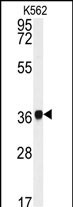
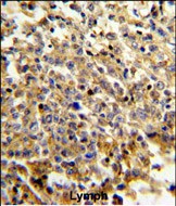
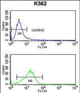
Application
| WB, IHC-P, FC, E |
|---|---|
| Primary Accession | Q15080 |
| Reactivity | Human |
| Host | Rabbit |
| Clonality | Polyclonal |
| Isotype | Rabbit IgG |
| Calculated MW | 39032 Da |
| Antigen Region | 260-289 aa |
| Gene ID | 4689 |
|---|---|
| Other Names | Neutrophil cytosol factor 4, NCF-4, Neutrophil NADPH oxidase factor 4, SH3 and PX domain-containing protein 4, p40-phox, p40phox, NCF4, SH3PXD4 |
| Target/Specificity | This NCF4 antibody is generated from rabbits immunized with a KLH conjugated synthetic peptide between 260-289 amino acids from the C-terminal region of human NCF4. |
| Dilution | WB~~1:1000 IHC-P~~1:100~500 FC~~1:10~50 E~~Use at an assay dependent concentration. |
| Format | Purified polyclonal antibody supplied in PBS with 0.09% (W/V) sodium azide. This antibody is prepared by Saturated Ammonium Sulfate (SAS) precipitation followed by dialysis against PBS. |
| Storage | Maintain refrigerated at 2-8°C for up to 2 weeks. For long term storage store at -20°C in small aliquots to prevent freeze-thaw cycles. |
| Precautions | NCF4 Antibody (C-term) is for research use only and not for use in diagnostic or therapeutic procedures. |
| Name | NCF4 (HGNC:7662) |
|---|---|
| Synonyms | SH3PXD4 |
| Function | Subunit of the phagocyte NADPH oxidase complex that mediates the transfer of electrons from cytosolic NADPH to O2 to produce the superoxide anion (O2(-)) (Probable). In the activated complex, electrons are first transferred from NADPH to flavin adenine dinucleotide (FAD) and subsequently transferred via two heme molecules to molecular oxygen, producing superoxide through an outer-sphere reaction (By similarity). Activation of the NADPH oxidase complex is initiated by the assembly of cytosolic subunits of the NADPH oxidase complex with the core NADPH oxidase complex to form a complex at the plasma membrane or phagosomal membrane (By similarity). This activation process is initiated by phosphorylation dependent binding of the cytosolic NCF1/p47-phox subunit to the C-terminus of CYBA/p22-phox (By similarity). |
| Cellular Location | Cytoplasm, cytosol. Endosome membrane; Peripheral membrane protein; Cytoplasmic side. Membrane; Peripheral membrane protein. Note=Translocates to the membrane upon activation by phorbol myristate acetate (PMA) |
| Tissue Location | Expression is restricted to hematopoietic cells. |
For Research Use Only. Not For Use In Diagnostic Procedures.
Provided below are standard protocols that you may find useful for product applications.
BACKGROUND
NCF4 is a cytosolic regulatory component of the superoxide-producing phagocyte NADPH-oxidase, a multicomponent enzyme system important for host defense. This protein is preferentially expressed in cells of myeloid lineage. It interacts primarily with neutrophil cytosolic factor 2(NCF2/p67-phox) to form a complex with neutrophil cytosolic factor 1 (NCF1/p47-phox), which further interacts with the small G protein RAC1 and translocates to the membrane upon cell stimulation. This complex then activates flavocytochrome b, the membrane-integrated catalytic core of the enzyme system. The PX domain of this protein can bind phospholipid products of the PI(3) kinase, which suggests its role in PI(3) kinase-mediated signaling events. The phosphorylation of this protein was found to negatively regulate the enzyme activity.
REFERENCES
Glas,J., Seiderer,J. Am. J. Gastroenterol. 104 (3), 665-672 (2009)
Honbou,K. Seikagaku 80 (8), 743-747 (2008)
Dusi,S., Donini,M. Biochem. J. 314 (PT 2), 409-412 (1996)
Leto,T.L. Proc. Natl. Acad. Sci. U.S.A. 91 (22), 10650-10654 (1994)
终于等到您。ABCEPTA(百远生物)抗体产品。
点击下方“我要评价 ”按钮提交您的反馈信息,您的反馈和评价是我们最宝贵的财富之一,
我们将在1-3个工作日内处理您的反馈信息。
如有疑问,联系:0512-88856768 tech-china@abcepta.com.
|
Anonymous
2016-08-24 09:20:44
1
2
3
4
5
|
Species tested
Human,Mouse,Rat
Application tested
WB
Organization tested
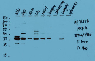
K562,Raji,HL-60,LO2,MCF-7,Lung(M),Spleen(M),Lung(R),Spleen(R)
Barcode encoding
Brief protocol
1. Block with 3% skim milk for 1 hour at room temperature.
2. Incubate overnight with Abgent primary antibody 1:1000 in 3% skim milk at 4℃
3. Wash 5*5 min with TBST.
4. Incubate with HRP-conjugated secondary antibody 1:5000 in 3% skim milk for 1 hour at room temperature.
5. Wash 5*5 min with TBST.
6. Incubate with ECL substrates and expose
|
|


