


 癌症的基本特征包括细胞增殖、血管生成、迁移、凋亡逃避机制和细胞永生等。找到癌症发生过程中这些通路的关键标记物和对应的抗体用于检测至关重要。
 为您推荐一个泛素化位点预测神器——泛素化分析工具,可以为您的蛋白的泛素化位点作出预测和评分。
 细胞自噬受体图形绘图工具为你的蛋白的细胞受体结合位点作出预测和评分,识别结合到自噬通路中的蛋白是非常重要的,便于让我们理解自噬在正常生理、病理过程中的作用,如发育、细胞分化、神经退化性疾病、压力条件下、感染和癌症。
LTK Antibody (N-term)
Purified Rabbit Polyclonal Antibody (Pab)
- 产品详情
- 实验流程
- 背景知识
Application
| WB, E |
|---|---|
| Primary Accession | P29376 |
| Reactivity | Human |
| Host | Rabbit |
| Clonality | Polyclonal |
| Isotype | Rabbit IgG |
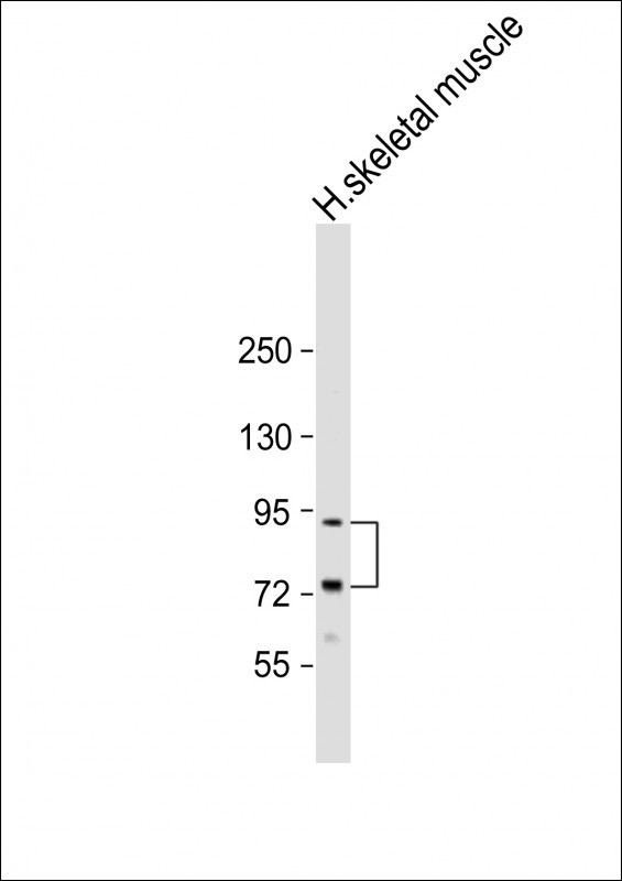
| Calculated MW | 91681 Da |
| Antigen Region | 42-72 aa |
| Gene ID | 4058 |
|---|---|
| Other Names | Leukocyte tyrosine kinase receptor, Protein tyrosine kinase 1, LTK, TYK1 |
| Target/Specificity | This LTK antibody is generated from rabbits immunized with a KLH conjugated synthetic peptide between 42-72 amino acids from the N-terminal region of human LTK. |
| Dilution | WB~~1:2000 E~~Use at an assay dependent concentration. |
| Format | Purified monoclonal antibody supplied in PBS with 0.09% (W/V) sodium azide. This antibody is purified through a protein G column, followed by dialysis against PBS. |
| Storage | Maintain refrigerated at 2-8°C for up to 2 weeks. For long term storage store at -20°C in small aliquots to prevent freeze-thaw cycles. |
| Precautions | LTK Antibody (N-term) is for research use only and not for use in diagnostic or therapeutic procedures. |
| Name | LTK {ECO:0000303|PubMed:1655406, ECO:0000312|HGNC:HGNC:6721} |
|---|---|
| Function | Receptor with a tyrosine-protein kinase activity (PubMed:10445845, PubMed:20548102, PubMed:30061385). Following activation by ALKAL1 or ALKAL2 ligands at the cell surface, transduces an extracellular signal into an intracellular response (PubMed:30061385, PubMed:34646012). Ligand-binding to the extracellular domain induces tyrosine kinase activation, leading to activation of the mitogen-activated protein kinase (MAPK) pathway (PubMed:20548102). Phosphorylates almost exclusively at the first tyrosine of the Y-x-x-x- Y-Y motif (By similarity). The exact function of this protein is not known; studies with chimeric proteins demonstrate its ability to promote growth and specifically neurite outgrowth, and cell survival (PubMed:18849880, PubMed:9223670). Involved in regulation of the secretory pathway involving endoplasmic reticulum (ER) export sites (ERESs) and ER to Golgi transport (PubMed:20548102). |
| Cellular Location | Cell membrane; Single-pass type I membrane protein |
| Tissue Location | Expressed in non-hematopoietic cell lines and T- and B-cell lines. |
For Research Use Only. Not For Use In Diagnostic Procedures.
Provided below are standard protocols that you may find useful for product applications.
BACKGROUND
Protein kinases are enzymes that transfer a phosphate group from a phosphate donor, generally the g phosphate of ATP, onto an acceptor amino acid in a substrate protein. By this basic mechanism, protein kinases mediate most of the signal transduction in eukaryotic cells, regulating cellular metabolism, transcription, cell cycle progression, cytoskeletal rearrangement and cell movement, apoptosis, and differentiation. With more than 500 gene products, the protein kinase family is one of the largest families of proteins in eukaryotes. The family has been classified in 8 major groups based on sequence comparison of their tyrosine (PTK) or serine/threonine (STK) kinase catalytic domains. The tyrosine kinase (TK) group is mainly involved in the regulation of cell-cell interactions such as differentiation, adhesion, motility and death. There are currently about 90 TK genes sequenced, 58 are of receptor protein TK (e.g. EGFR, EPH, FGFR, PDGFR, TRK, and VEGFR families), and 32 of cytosolic TK (e.g. ABL, FAK, JAK, and SRC families).
REFERENCES
Toyoshima, H., et al., Proc. Natl. Acad. Sci. U.S.A. 90(12):5404-5408 (1993).
Krolewski, J.J., et al., EMBO J. 10(10):2911-2919 (1991).
Maru, Y., et al., Oncogene Res. 5(3):199-204 (1990).
终于等到您。ABCEPTA(百远生物)抗体产品。
点击下方“我要评价 ”按钮提交您的反馈信息,您的反馈和评价是我们最宝贵的财富之一,
我们将在1-3个工作日内处理您的反馈信息。
如有疑问,联系:0512-88856768 tech-china@abcepta.com.


