


 癌症的基本特征包括细胞增殖、血管生成、迁移、凋亡逃避机制和细胞永生等。找到癌症发生过程中这些通路的关键标记物和对应的抗体用于检测至关重要。
 为您推荐一个泛素化位点预测神器——泛素化分析工具,可以为您的蛋白的泛素化位点作出预测和评分。
 细胞自噬受体图形绘图工具为你的蛋白的细胞受体结合位点作出预测和评分,识别结合到自噬通路中的蛋白是非常重要的,便于让我们理解自噬在正常生理、病理过程中的作用,如发育、细胞分化、神经退化性疾病、压力条件下、感染和癌症。
TXK Antibody (N-term)
Purified Rabbit Polyclonal Antibody (Pab)
- 产品详情
- 实验流程
- 背景知识
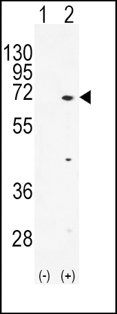
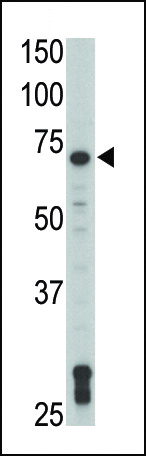
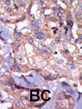
Application
| WB, IHC-P, E |
|---|---|
| Primary Accession | P42681 |
| Reactivity | Human |
| Host | Rabbit |
| Clonality | Polyclonal |
| Isotype | Rabbit IgG |
| Calculated MW | 61258 Da |
| Antigen Region | 22-52 aa |
| Gene ID | 7294 |
|---|---|
| Other Names | Tyrosine-protein kinase TXK, Protein-tyrosine kinase 4, Resting lymphocyte kinase, TXK, PTK4, RLK |
| Target/Specificity | This TXK antibody is generated from rabbits immunized with a KLH conjugated synthetic peptide between 22-52 amino acids from the N-terminal region of human TXK. |
| Dilution | WB~~1:1000 IHC-P~~1:100~500 E~~Use at an assay dependent concentration. |
| Format | Purified polyclonal antibody supplied in PBS with 0.09% (W/V) sodium azide. This antibody is prepared by Saturated Ammonium Sulfate (SAS) precipitation followed by dialysis against PBS. |
| Storage | Maintain refrigerated at 2-8°C for up to 2 weeks. For long term storage store at -20°C in small aliquots to prevent freeze-thaw cycles. |
| Precautions | TXK Antibody (N-term) is for research use only and not for use in diagnostic or therapeutic procedures. |
| Name | TXK |
|---|---|
| Synonyms | PTK4, RLK |
| Function | Non-receptor tyrosine kinase that plays a redundant role with ITK in regulation of the adaptive immune response. Regulates the development, function and differentiation of conventional T-cells and nonconventional NKT-cells. When antigen presenting cells (APC) activate T-cell receptor (TCR), a series of phosphorylation leads to the recruitment of TXK to the cell membrane, where it is phosphorylated at Tyr-420. Phosphorylation leads to TXK full activation. Also contributes to signaling from many receptors and participates in multiple downstream pathways, including regulation of the actin cytoskeleton. Like ITK, can phosphorylate PLCG1, leading to its localization in lipid rafts and activation, followed by subsequent cleavage of its substrates. In turn, the endoplasmic reticulum releases calcium in the cytoplasm and the nuclear activator of activated T-cells (NFAT) translocates into the nucleus to perform its transcriptional duty. Plays a role in the positive regulation of IFNG transcription in T- helper 1 cells as part of an IFNG promoter-binding complex with PARP1 and EEF1A1 (PubMed:11859127, PubMed:17177976). Within the complex, phosphorylates both PARP1 and EEF1A1 (PubMed:17177976). Also phosphorylates key sites in LCP2 leading to the up-regulation of Th1 preferred cytokine IL-2. Phosphorylates 'Tyr-201' of CTLA4 which leads to the association of PI-3 kinase with the CTLA4 receptor. |
| Cellular Location | Cytoplasm. Nucleus. Cell membrane; Peripheral membrane protein. Note=Localizes in the vicinity of cell surface receptors in the plasma membrane after receptor stimulation Translocates into the nucleus and enhances IFN-gamma gene transcription in T-cells |
| Tissue Location | Expressed in T-cells and some myeloid cell lines. Expressed in Th1/Th0 cells with IFN-gamma-producing potential |
For Research Use Only. Not For Use In Diagnostic Procedures.
Provided below are standard protocols that you may find useful for product applications.
BACKGROUND
Protein kinases are enzymes that transfer a phosphate group from a phosphate donor, generally the g phosphate of ATP, onto an acceptor amino acid in a substrate protein. By this basic mechanism, protein kinases mediate most of the signal transduction in eukaryotic cells, regulating cellular metabolism, transcription, cell cycle progression, cytoskeletal rearrangement and cell movement, apoptosis, and differentiation. With more than 500 gene products, the protein kinase family is one of the largest families of proteins in eukaryotes. The family has been classified in 8 major groups based on sequence comparison of their tyrosine (PTK) or serine/threonine (STK) kinase catalytic domains. The STE group (homologs of yeast Sterile 7, 11, 20 kinases) consists of 50 kinases related to the mitogen-activated protein kinase (MAPK) cascade families (Ste7/MAP2K, Ste11/MAP3K, and Ste20/MAP4K). MAP kinase cascades, consisting of a MAPK and one or more upstream regulatory kinases (MAPKKs) have been best characterized in the yeast pheromone response pathway. Pheromones bind to Ste cell surface receptors and activate yeast MAPK pathway.
REFERENCES
Takeba, Y., et al., J. Immunol. 168(5):2365-2370 (2002). Spritz, R.A., et al., Genomics 22(2):431-436 (1994). Haire, R.N., et al., Hum. Mol. Genet. 3(6):897-901 (1994).
终于等到您。ABCEPTA(百远生物)抗体产品。
点击下方“我要评价 ”按钮提交您的反馈信息,您的反馈和评价是我们最宝贵的财富之一,
我们将在1-3个工作日内处理您的反馈信息。
如有疑问,联系:0512-88856768 tech-china@abcepta.com.


