


 癌症的基本特征包括细胞增殖、血管生成、迁移、凋亡逃避机制和细胞永生等。找到癌症发生过程中这些通路的关键标记物和对应的抗体用于检测至关重要。
 为您推荐一个泛素化位点预测神器——泛素化分析工具,可以为您的蛋白的泛素化位点作出预测和评分。
 细胞自噬受体图形绘图工具为你的蛋白的细胞受体结合位点作出预测和评分,识别结合到自噬通路中的蛋白是非常重要的,便于让我们理解自噬在正常生理、病理过程中的作用,如发育、细胞分化、神经退化性疾病、压力条件下、感染和癌症。
Heparanase 1 Rabbit mAb
- 产品详情
- 实验流程
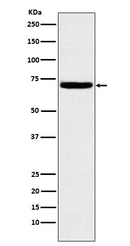
Application
| WB, IP |
|---|---|
| Primary Accession | Q9Y251 |
| Reactivity | Rat, Human, Mouse |
| Host | Rabbit |
| Clonality | Monoclonal Antibody |
| Isotype | IgG |
| Conjugate | Unconjugated |
| Immunogen | A synthesized peptide derived from human Heparanase 1 |
| Purification | Affinity Chromatography |
| Calculated MW | 61149 Da |
| Gene ID | 10855 |
|---|---|
| Other Names | HPSE |
| Dilution | WB~~1/500-1/1000 IP~~N/A |
| Format | Liquid in 10mM PBS, pH 7.4, 150mM sodium chloride, 0.05% BSA, 0.02% sodium azide and 50% glycerol. |
| Storage | Store at 4°C short term. Aliquot and store at -20°C long term. Avoid freeze/thaw cycles. |
| Name | HPSE |
|---|---|
| Synonyms | HEP, HPA, HPA1, HPR1, HPSE1, HSE1 |
| Function | Endoglycosidase that cleaves heparan sulfate proteoglycans (HSPGs) into heparan sulfate side chains and core proteoglycans. Participates in extracellular matrix (ECM) degradation and remodeling. Selectively cleaves the linkage between a glucuronic acid unit and an N-sulfo glucosamine unit carrying either a 3-O-sulfo or a 6-O-sulfo group. Can also cleave the linkage between a glucuronic acid unit and an N-sulfo glucosamine unit carrying a 2-O-sulfo group, but not linkages between a glucuronic acid unit and a 2-O-sulfated iduronic acid moiety. It is essentially inactive at neutral pH but becomes active under acidic conditions such as during tumor invasion and in inflammatory processes. Facilitates cell migration associated with metastasis, wound healing and inflammation. Enhances shedding of syndecans, and increases endothelial invasion and angiogenesis in myelomas. Acts as a procoagulant by increasing the generation of activation factor X in the presence of tissue factor and activation factor VII. Increases cell adhesion to the extracellular matrix (ECM), independent of its enzymatic activity. Induces AKT1/PKB phosphorylation via lipid rafts increasing cell mobility and invasion. Heparin increases this AKT1/PKB activation. Regulates osteogenesis. Enhances angiogenesis through up-regulation of SRC-mediated activation of VEGF. Implicated in hair follicle inner root sheath differentiation and hair homeostasis. |
| Cellular Location | Lysosome membrane; Peripheral membrane protein. Secreted. Nucleus. Note=Proheparanase is secreted via vesicles of the Golgi. Interacts with cell membrane heparan sulfate proteoglycans (HSPGs). Endocytosed and accumulates in endosomes. Transferred to lysosomes where it is proteolytically cleaved to produce the active enzyme. Under certain stimuli, transferred to the cell surface Associates with lipid rafts. Colocalizes with SDC1 in endosomal/lysosomal vesicles. Accumulates in perinuclear lysosomal vesicles. Heparin retains proheparanase in the extracellular medium (By similarity). |
| Tissue Location | Highly expressed in placenta and spleen and weakly expressed in lymph node, thymus, peripheral blood leukocytes, bone marrow, endothelial cells, fetal liver and tumor tissues. Also expressed in hair follicles, specifically in both Henle's and Huxley's layers of inner the root sheath (IRS) at anagen phase |
Research Areas
For Research Use Only. Not For Use In Diagnostic Procedures.
Application Protocols
Provided below are standard protocols that you may find useful for product applications.
终于等到您。ABCEPTA(百远生物)抗体产品。
点击下方“我要评价 ”按钮提交您的反馈信息,您的反馈和评价是我们最宝贵的财富之一,
我们将在1-3个工作日内处理您的反馈信息。
如有疑问,联系:0512-88856768 tech-china@abcepta.com.
¥ 1,500.00
Cat# AP77890


