


 癌症的基本特征包括细胞增殖、血管生成、迁移、凋亡逃避机制和细胞永生等。找到癌症发生过程中这些通路的关键标记物和对应的抗体用于检测至关重要。
 为您推荐一个泛素化位点预测神器——泛素化分析工具,可以为您的蛋白的泛素化位点作出预测和评分。
 细胞自噬受体图形绘图工具为你的蛋白的细胞受体结合位点作出预测和评分,识别结合到自噬通路中的蛋白是非常重要的,便于让我们理解自噬在正常生理、病理过程中的作用,如发育、细胞分化、神经退化性疾病、压力条件下、感染和癌症。
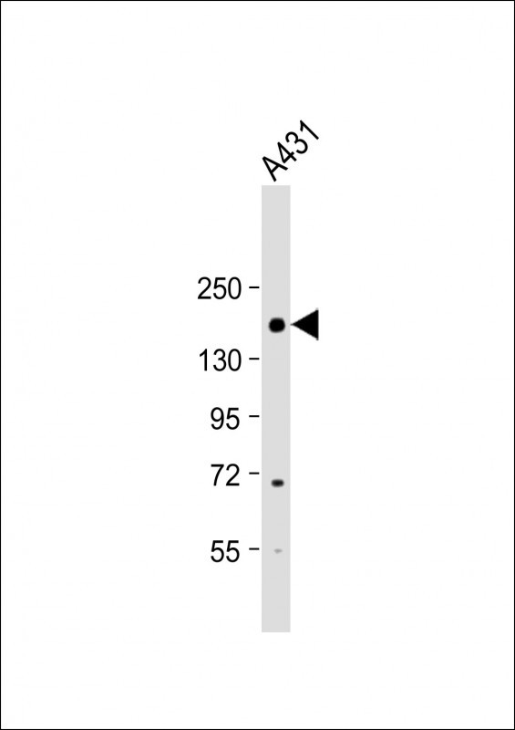
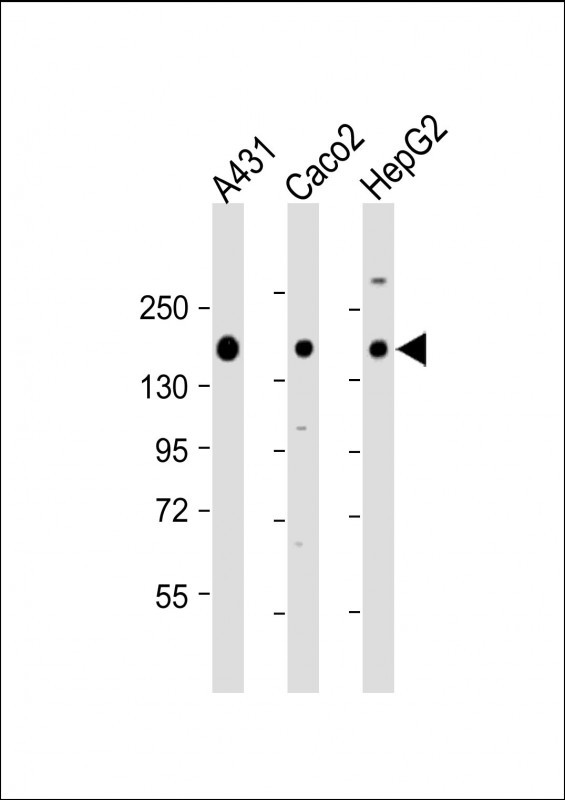
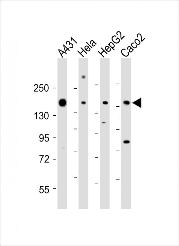
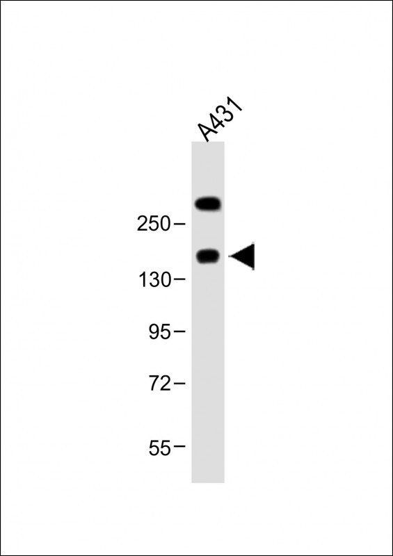
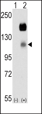
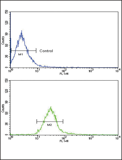
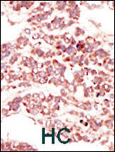
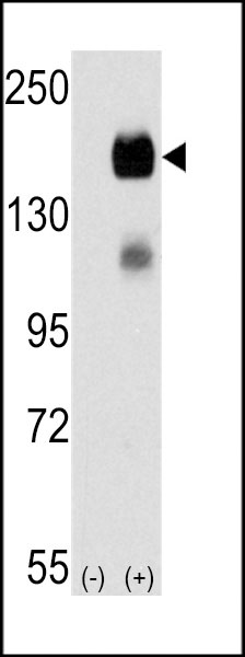
CCK4 (PTK7) Antibody (N-term)
Purified Rabbit Polyclonal Antibody (Pab)
- 产品详情
- 文献引用 : 1
- 实验流程
- 背景知识
Application
| FC, IHC-P, WB, E |
|---|---|
| Primary Accession | Q13308 |
| Reactivity | Human, Mouse |
| Host | Rabbit |
| Clonality | Polyclonal |
| Isotype | Rabbit IgG |
| Calculated MW | 118392 Da |
| Antigen Region | 21-52 aa |
| Gene ID | 5754 |
|---|---|
| Other Names | Inactive tyrosine-protein kinase 7, Colon carcinoma kinase 4, CCK-4, Protein-tyrosine kinase 7, Pseudo tyrosine kinase receptor 7, Tyrosine-protein kinase-like 7, PTK7, CCK4 |
| Target/Specificity | This CCK4 (PTK7) antibody is generated from rabbits immunized with a KLH conjugated synthetic peptide between 21-52 amino acids from the N-terminal region of human CCK4 (PTK7). |
| Dilution | FC~~1:10~50 IHC-P~~1:100~500 WB~~1:1000 E~~Use at an assay dependent concentration. |
| Format | Purified polyclonal antibody supplied in PBS with 0.09% (W/V) sodium azide. This antibody is purified through a protein A column, followed by peptide affinity purification. |
| Storage | Maintain refrigerated at 2-8°C for up to 2 weeks. For long term storage store at -20°C in small aliquots to prevent freeze-thaw cycles. |
| Precautions | CCK4 (PTK7) Antibody (N-term) is for research use only and not for use in diagnostic or therapeutic procedures. |
| Name | PTK7 |
|---|---|
| Synonyms | CCK4 |
| Function | Inactive tyrosine kinase involved in Wnt signaling pathway. Component of both the non-canonical (also known as the Wnt/planar cell polarity signaling) and the canonical Wnt signaling pathway. Functions in cell adhesion, cell migration, cell polarity, proliferation, actin cytoskeleton reorganization and apoptosis. Has a role in embryogenesis, epithelial tissue organization and angiogenesis. |
| Cellular Location | Membrane; Single- pass type I membrane protein. Cell junction. Note=Colocalizes with MMP14 at cell junctions. Also localizes at the leading edge of migrating cells |
| Tissue Location | Highly expressed in lung, liver, pancreas, kidney, placenta and melanocytes. Weakly expressed in thyroid gland, ovary, brain, heart and skeletal muscle. Also expressed in erythroleukemia cells. But not expressed in colon |
For Research Use Only. Not For Use In Diagnostic Procedures.

Provided below are standard protocols that you may find useful for product applications.
BACKGROUND
CCK4 may function as a cell adhesion molecule. Although it belongs to the insuline receptor subfamily of the Tyr protein kinases, it likely lacks the catalytic activity of a tyrosine kinase. It may be connected to the pathophysiology of colon carcinomas and/or may represent a tumor progression marker. This Type I membrane protein is highly expressed in lung, liver, pancreas, kidney, placenta and melanocytes, but weakly expressed in thyroid gland, ovary, brain, heart and skeletal muscle, and not in colon. It is also expressed in erythroleukemia cells.
REFERENCES
Zhang, H., et al., Nat. Biotechnol. 21(6):660-666 (2003).
Park, S.K., et al., J. Biochem. 119(2):235-239 (1996).
Mossie, K., et al., Oncogene 11(10):2179-2184 (1995).
终于等到您。ABCEPTA(百远生物)抗体产品。
点击下方“我要评价 ”按钮提交您的反馈信息,您的反馈和评价是我们最宝贵的财富之一,
我们将在1-3个工作日内处理您的反馈信息。
如有疑问,联系:0512-88856768 tech-china@abcepta.com.


