


 癌症的基本特征包括细胞增殖、血管生成、迁移、凋亡逃避机制和细胞永生等。找到癌症发生过程中这些通路的关键标记物和对应的抗体用于检测至关重要。
 为您推荐一个泛素化位点预测神器——泛素化分析工具,可以为您的蛋白的泛素化位点作出预测和评分。
 细胞自噬受体图形绘图工具为你的蛋白的细胞受体结合位点作出预测和评分,识别结合到自噬通路中的蛋白是非常重要的,便于让我们理解自噬在正常生理、病理过程中的作用,如发育、细胞分化、神经退化性疾病、压力条件下、感染和癌症。
PAK2 Antibody (N-term)
Purified Rabbit Polyclonal Antibody (Pab)

- 产品详情
- 实验流程
- 背景知识
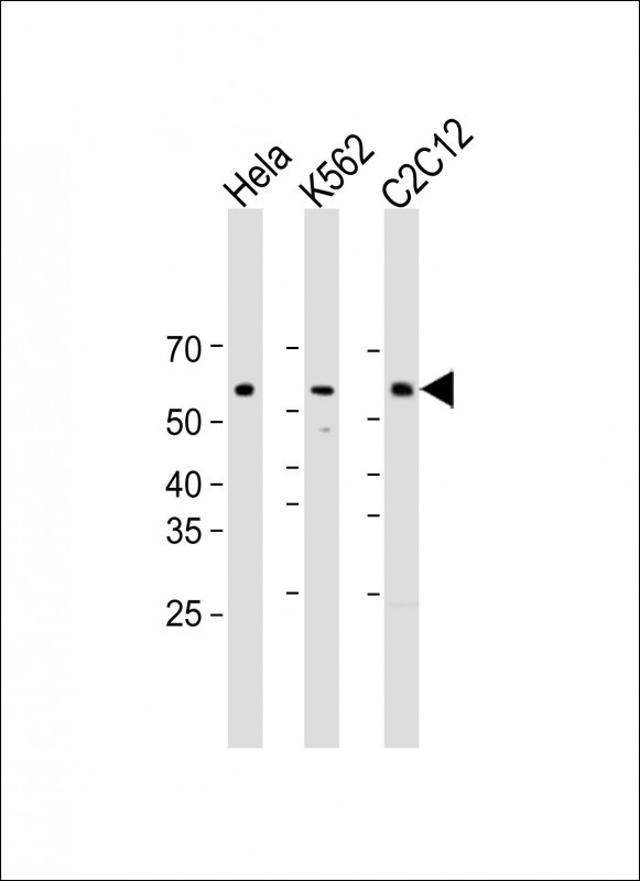
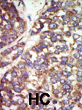
Application
| WB, IHC-P, E |
|---|---|
| Primary Accession | Q13177 |
| Reactivity | Human |
| Host | Rabbit |
| Clonality | Polyclonal |
| Isotype | Rabbit IgG |
| Calculated MW | 58043 Da |
| Antigen Region | 192-222 aa |
| Gene ID | 5062 |
|---|---|
| Other Names | Serine/threonine-protein kinase PAK 2, Gamma-PAK, PAK65, S6/H4 kinase, p21-activated kinase 2, PAK-2, p58, PAK-2p27, p27, PAK-2p34, p34, C-t-PAK2, PAK2 |
| Target/Specificity | This PAK2 antibody is generated from rabbits immunized with a KLH conjugated synthetic peptide between 192-222 amino acids from the N-terminal region of human PAK2. |
| Dilution | WB~~1:1000 IHC-P~~1:100~500 E~~Use at an assay dependent concentration. |
| Format | Purified monoclonal antibody supplied in PBS with 0.09% (W/V) sodium azide. This antibody is purified through a protein G column, followed by dialysis against PBS. |
| Storage | Maintain refrigerated at 2-8°C for up to 2 weeks. For long term storage store at -20°C in small aliquots to prevent freeze-thaw cycles. |
| Precautions | PAK2 Antibody (N-term) is for research use only and not for use in diagnostic or therapeutic procedures. |
| Name | PAK2 |
|---|---|
| Function | Serine/threonine protein kinase that plays a role in a variety of different signaling pathways including cytoskeleton regulation, cell motility, cell cycle progression, apoptosis or proliferation (PubMed:12853446, PubMed:16617111, PubMed:19273597, PubMed:19923322, PubMed:33693784, PubMed:7744004, PubMed:9171063). Acts as a downstream effector of the small GTPases CDC42 and RAC1 (PubMed:7744004). Activation by the binding of active CDC42 and RAC1 results in a conformational change and a subsequent autophosphorylation on several serine and/or threonine residues (PubMed:7744004). Full- length PAK2 stimulates cell survival and cell growth (PubMed:7744004). Phosphorylates MAPK4 and MAPK6 and activates the downstream target MAPKAPK5, a regulator of F-actin polymerization and cell migration (PubMed:21317288). Phosphorylates JUN and plays an important role in EGF-induced cell proliferation (PubMed:21177766). Phosphorylates many other substrates including histone H4 to promote assembly of H3.3 and H4 into nucleosomes, BAD, ribosomal protein S6, or MBP (PubMed:21724829). Phosphorylates CASP7, thereby preventing its activity (PubMed:21555521, PubMed:27889207). Additionally, associates with ARHGEF7 and GIT1 to perform kinase-independent functions such as spindle orientation control during mitosis (PubMed:19273597, PubMed:19923322). On the other hand, apoptotic stimuli such as DNA damage lead to caspase-mediated cleavage of PAK2, generating PAK-2p34, an active p34 fragment that translocates to the nucleus and promotes cellular apoptosis involving the JNK signaling pathway (PubMed:12853446, PubMed:16617111, PubMed:9171063). Caspase-activated PAK2 phosphorylates MKNK1 and reduces cellular translation (PubMed:15234964). |
| Cellular Location | [Serine/threonine-protein kinase PAK 2]: Cytoplasm Nucleus Note=MYO18A mediates the cellular distribution of the PAK2-ARHGEF7-GIT1 complex to the inner surface of the cell membrane |
| Tissue Location | Ubiquitously expressed. Higher levels seen in skeletal muscle, ovary, thymus and spleen |
For Research Use Only. Not For Use In Diagnostic Procedures.
Provided below are standard protocols that you may find useful for product applications.
BACKGROUND
PAK2, a member of the STE20 subfamily of Ser/Thr protein kinases, acts on a variety of targets. It phosphorylates ribosomal protein S6, histone H4 and myelin basic protein. PAK2 interacts tightly with GTP-bound but not GDP-bound CDC42/p21 and RAC1. Expression is ubiquitous, with higher levels seen in skeletal muscle, ovary, thymus and spleen. PAK2 is autophosphorylated when activated by CDC42/p21. The protein structure contains 1 CRIB domain.
REFERENCES
Benner, G.E., et al., J. Biol. Chem. 270(36):21121-21128 (1995).
Martin, G.A., et al., EMBO J. 14(9):1970-1978 (1995).
Martin, G.A., et al., EMBO J. 14 (17), 4385 (1995).
终于等到您。ABCEPTA(百远生物)抗体产品。
点击下方“我要评价 ”按钮提交您的反馈信息,您的反馈和评价是我们最宝贵的财富之一,
我们将在1-3个工作日内处理您的反馈信息。
如有疑问,联系:0512-88856768 tech-china@abcepta.com.


