


 癌症的基本特征包括细胞增殖、血管生成、迁移、凋亡逃避机制和细胞永生等。找到癌症发生过程中这些通路的关键标记物和对应的抗体用于检测至关重要。
 为您推荐一个泛素化位点预测神器——泛素化分析工具,可以为您的蛋白的泛素化位点作出预测和评分。
 细胞自噬受体图形绘图工具为你的蛋白的细胞受体结合位点作出预测和评分,识别结合到自噬通路中的蛋白是非常重要的,便于让我们理解自噬在正常生理、病理过程中的作用,如发育、细胞分化、神经退化性疾病、压力条件下、感染和癌症。
PKD2 Antibody (C-term)
Purified Rabbit Polyclonal Antibody (Pab)
- 产品详情
- 实验流程
- 背景知识
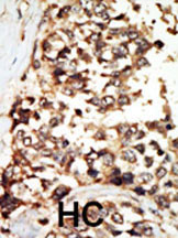
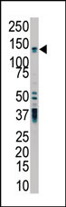
Application
| IHC-P, WB, E |
|---|---|
| Primary Accession | Q13563 |
| Reactivity | Human |
| Host | Rabbit |
| Clonality | Polyclonal |
| Isotype | Rabbit IgG |
| Calculated MW | 109691 Da |
| Antigen Region | 937-968 aa |
| Gene ID | 5311 |
|---|---|
| Other Names | Polycystin-2, Autosomal dominant polycystic kidney disease type II protein, Polycystic kidney disease 2 protein, Polycystwin, R48321, PKD2 |
| Target/Specificity | This PKD2 antibody is generated from rabbits immunized with a KLH conjugated synthetic peptide between 937-968 amino acids from the C-terminal region of human PKD2. |
| Dilution | IHC-P~~1:100~500 WB~~1:1000 E~~Use at an assay dependent concentration. |
| Format | Purified monoclonal antibody supplied in PBS with 0.09% (W/V) sodium azide. This antibody is purified through a protein G column, followed by dialysis against PBS. |
| Storage | Maintain refrigerated at 2-8°C for up to 2 weeks. For long term storage store at -20°C in small aliquots to prevent freeze-thaw cycles. |
| Precautions | PKD2 Antibody (C-term) is for research use only and not for use in diagnostic or therapeutic procedures. |
| Name | PKD2 (HGNC:9009) |
|---|---|
| Function | Forms a nonselective cation channel (PubMed:11854751, PubMed:11991947, PubMed:15692563, PubMed:26269590, PubMed:27071085, PubMed:31441214, PubMed:39009345). Can function as a homotetrameric ion channel or can form heteromer with PKD1 (PubMed:31441214, PubMed:33164752). Displays distinct function depending on its subcellular localization and regulation by its binding partners (PubMed:11854751, PubMed:11991947, PubMed:27214281, PubMed:29899465). In primary cilium functions as a cation channel, with a preference for monovalent cations over divalent cations that allows K(+), Na(+) and Ca(2+) influx, with low selectivity for Ca(2+) (PubMed:27071085). Involved in fluid-flow mechanosensation by the primary cilium in renal epithelium (By similarity). In the endoplasmic reticulum, likely functions as a K(+) channel to facilitate Ca(2+) release (By similarity). The heterotetrameric PKD1/PKD2 channel has higher Ca(2+) permeability than homomeric PKD2 channel and acts as a primarily Ca(2+)-permeable channel (PubMed:31441214). Interacts with and acts as a regulator of a number of other channels, such as TRPV4, TRPC1, IP3R, RYR2, ultimately further affecting intracellular signaling, to modulate intracellular Ca(2+) signaling (PubMed:11854751, PubMed:11991947, PubMed:27214281, PubMed:29899465). Together with TRPV4, forms mechano- and thermosensitive channels in cilium (PubMed:18695040). In cardiomyocytes, PKD2 modulates Ca(2+) release from stimulated RYR2 receptors through direct association (By similarity). Also involved in left-right axis specification via its role in sensing nodal flow; forms a complex with PKD1L1 in cilia to facilitate flow detection in left- right patterning (By similarity). Acts as a regulator of cilium length together with PKD1 (By similarity). Mediates systemic blood pressure and contributes to the myogenic response in cerebral arteries though vasoconstriction (By similarity). |
| Cellular Location | Cell projection, cilium membrane; Multi-pass membrane protein. Endoplasmic reticulum membrane; Multi-pass membrane protein. Cell membrane; Multi-pass membrane protein. Basolateral cell membrane. Cytoplasmic vesicle membrane. Golgi apparatus {ECO:0000250|UniProtKB:O35245}. Vesicle Secreted, extracellular exosome Note=PKD2 localization to the plasma and ciliary membranes requires PKD1. PKD1:PKD2 interaction is required to reach the Golgi apparatus form endoplasmic reticulum and then traffic to the cilia (By similarity). Retained in the endoplasmic reticulum by interaction with PACS1 and PACS2 (PubMed:15692563). Detected on kidney tubule basolateral membranes and basal cytoplasmic vesicles (PubMed:10770959) Cell surface and cilium localization requires GANAB (PubMed:27259053) Detected on migrasomes and on extracellular exosomes in urine (PubMed:21406692). Preferentially localized to the dorsal side of immotile cilia (By similarity). {ECO:0000250|UniProtKB:O35245, ECO:0000269|PubMed:15692563, ECO:0000269|PubMed:21406692, ECO:0000269|PubMed:27259053} |
| Tissue Location | Detected in fetal and adult kidney (PubMed:10770959). Detected at the thick ascending limb of the loop of Henle, at distal tubules, including the distal convoluted tubule and cortical collecting tubules, with weak staining of the collecting duct (PubMed:10770959). Detected on placenta syncytiotrophoblasts (at protein level) (PubMed:26269590). Strongly expressed in ovary, fetal and adult kidney, testis, and small intestine. Not detected in peripheral leukocytes. |
For Research Use Only. Not For Use In Diagnostic Procedures.
Provided below are standard protocols that you may find useful for product applications.
BACKGROUND
Involved in fluid-flow mechanosensation by the primary cilium in renal epithelium. PKD1 and PKD2 may function through a common signaling pathway that is necessary for normal tubulogenesis. Acts as a regulator of cilium length, together with PKD1. The dynamic control of cilium length is essential in the regulation of mechanotransductive signaling. The cilium length response creates a negative feedback loop whereby fluid shear-mediated deflection of the primary cilium, which decreases intracellular cAMP, leads to cilium shortening and thus decreases flow-induced signaling. Functions as a calcium permeable cation channel.
终于等到您。ABCEPTA(百远生物)抗体产品。
点击下方“我要评价 ”按钮提交您的反馈信息,您的反馈和评价是我们最宝贵的财富之一,
我们将在1-3个工作日内处理您的反馈信息。
如有疑问,联系:0512-88856768 tech-china@abcepta.com.


