


 癌症的基本特征包括细胞增殖、血管生成、迁移、凋亡逃避机制和细胞永生等。找到癌症发生过程中这些通路的关键标记物和对应的抗体用于检测至关重要。
 为您推荐一个泛素化位点预测神器——泛素化分析工具,可以为您的蛋白的泛素化位点作出预测和评分。
 细胞自噬受体图形绘图工具为你的蛋白的细胞受体结合位点作出预测和评分,识别结合到自噬通路中的蛋白是非常重要的,便于让我们理解自噬在正常生理、病理过程中的作用,如发育、细胞分化、神经退化性疾病、压力条件下、感染和癌症。
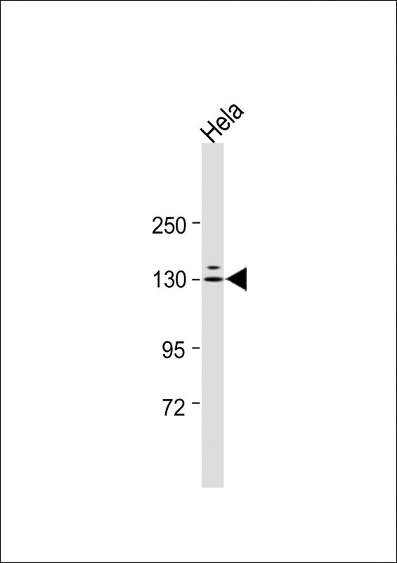
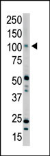
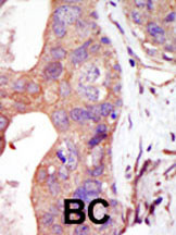
PRK2 Antibody (C-term)
Purified Rabbit Polyclonal Antibody (Pab)
- 产品详情
- 实验流程
- 背景知识
Application
| WB, IHC-P, E |
|---|---|
| Primary Accession | Q16513 |
| Reactivity | Human, Rat, Mouse |
| Host | Rabbit |
| Clonality | Polyclonal |
| Isotype | Rabbit IgG |
| Calculated MW | 112035 Da |
| Antigen Region | 627-658 aa |
| Gene ID | 5586 |
|---|---|
| Other Names | Serine/threonine-protein kinase N2, PKN gamma, Protein kinase C-like 2, Protein-kinase C-related kinase 2, PKN2, PRK2, PRKCL2 |
| Target/Specificity | This PRK2 antibody is generated from rabbits immunized with a KLH conjugated synthetic peptide between 627-658 amino acids from the C-terminal region of human PRK2. |
| Dilution | WB~~1:1000 IHC-P~~1:100~500 E~~Use at an assay dependent concentration. |
| Format | Purified polyclonal antibody supplied in PBS with 0.05% (V/V) Proclin 300. This antibody is prepared by Saturated Ammonium Sulfate (SAS) precipitation followed by dialysis against PBS. |
| Storage | Maintain refrigerated at 2-8°C for up to 2 weeks. For long term storage store at -20°C in small aliquots to prevent freeze-thaw cycles. |
| Precautions | PRK2 Antibody (C-term) is for research use only and not for use in diagnostic or therapeutic procedures. |
| Name | PKN2 |
|---|---|
| Synonyms | PRK2, PRKCL2 |
| Function | PKC-related serine/threonine-protein kinase and Rho/Rac effector protein that participates in specific signal transduction responses in the cell. Plays a role in the regulation of cell cycle progression, actin cytoskeleton assembly, cell migration, cell adhesion, tumor cell invasion and transcription activation signaling processes. Phosphorylates CTTN in hyaluronan-induced astrocytes and hence decreases CTTN ability to associate with filamentous actin. Phosphorylates HDAC5, therefore lead to impair HDAC5 import. Direct RhoA target required for the regulation of the maturation of primordial junctions into apical junction formation in bronchial epithelial cells. Required for G2/M phases of the cell cycle progression and abscission during cytokinesis in a ECT2-dependent manner. Stimulates FYN kinase activity that is required for establishment of skin cell-cell adhesion during keratinocytes differentiation. Regulates epithelial bladder cells speed and direction of movement during cell migration and tumor cell invasion. Inhibits Akt pro-survival-induced kinase activity. Mediates Rho protein-induced transcriptional activation via the c-fos serum response factor (SRF). Involved in the negative regulation of ciliogenesis (PubMed:27104747). |
| Cellular Location | Cytoplasm. Nucleus Membrane {ECO:0000250|UniProtKB:Q8BWW9}. Cell projection, lamellipodium. Cytoplasm, cytoskeleton. Cleavage furrow. Midbody Cell junction. Note=Colocalizes with PTPN13 in lamellipodia-like structures, regions of large actin turnover. Accumulates during telophase at the cleavage furrow and concentrates finally around the midbody in cytokinesis. Recruited to nascent cell-cell contacts at the apical surface of cells. In the course of viral infection, colocalizes with HCV NS5B at perinuclear region in the cytoplasm. |
| Tissue Location | Ubiquitous. Expressed in numerous tumor cell lines, especially in bladder tumor cells. |
For Research Use Only. Not For Use In Diagnostic Procedures.
Provided below are standard protocols that you may find useful for product applications.
BACKGROUND
Protein kinases are enzymes that transfer a phosphate group from a phosphate donor, generally the g phosphate of ATP, onto an acceptor amino acid in a substrate protein. By this basic mechanism, protein kinases mediate most of the signal transduction in eukaryotic cells, regulating cellular metabolism, transcription, cell cycle progression, cytoskeletal rearrangement and cell movement, apoptosis, and differentiation. With more than 500 gene products, the protein kinase family is one of the largest families of proteins in eukaryotes. The family has been classified in 8 major groups based on sequence comparison of their tyrosine (PTK) or serine/threonine (STK) kinase catalytic domains. The AGC kinase group consists of 63 kinases including the cyclic nucleotide-regulated protein kinase (PKA & PKG) family, the diacylglycerol-activated/phospholipid-dependent protein kinase C (PKC) family, the related to PKA and PKC (RAC/Akt) protein kinase family, the kinases that phosphorylate G protein-coupled receptors family (ARK), and the kinases that phosphorylate ribosomal protein S6 family (RSK). The calcium/calmodulin-dependent kinase (CAMK) group consists of 75 kinases regulated by Ca2+/CaM and close relative family (CAMK, CAMKL, DAPK, MAPKAPK).
REFERENCES
Yu, W., et al., J. Biol. Chem. 272(15):10030-10034 (1997).
Palmer, R.H., et al., FEBS Lett. 356(1):5-8 (1994).
Palmer, R.H., et al., Eur. J. Biochem. 227 (1-2), 344-351 (1995).
终于等到您。ABCEPTA(百远生物)抗体产品。
点击下方“我要评价 ”按钮提交您的反馈信息,您的反馈和评价是我们最宝贵的财富之一,
我们将在1-3个工作日内处理您的反馈信息。
如有疑问,联系:0512-88856768 tech-china@abcepta.com.


