


 癌症的基本特征包括细胞增殖、血管生成、迁移、凋亡逃避机制和细胞永生等。找到癌症发生过程中这些通路的关键标记物和对应的抗体用于检测至关重要。
 为您推荐一个泛素化位点预测神器——泛素化分析工具,可以为您的蛋白的泛素化位点作出预测和评分。
 细胞自噬受体图形绘图工具为你的蛋白的细胞受体结合位点作出预测和评分,识别结合到自噬通路中的蛋白是非常重要的,便于让我们理解自噬在正常生理、病理过程中的作用,如发育、细胞分化、神经退化性疾病、压力条件下、感染和癌症。
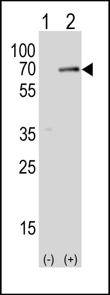
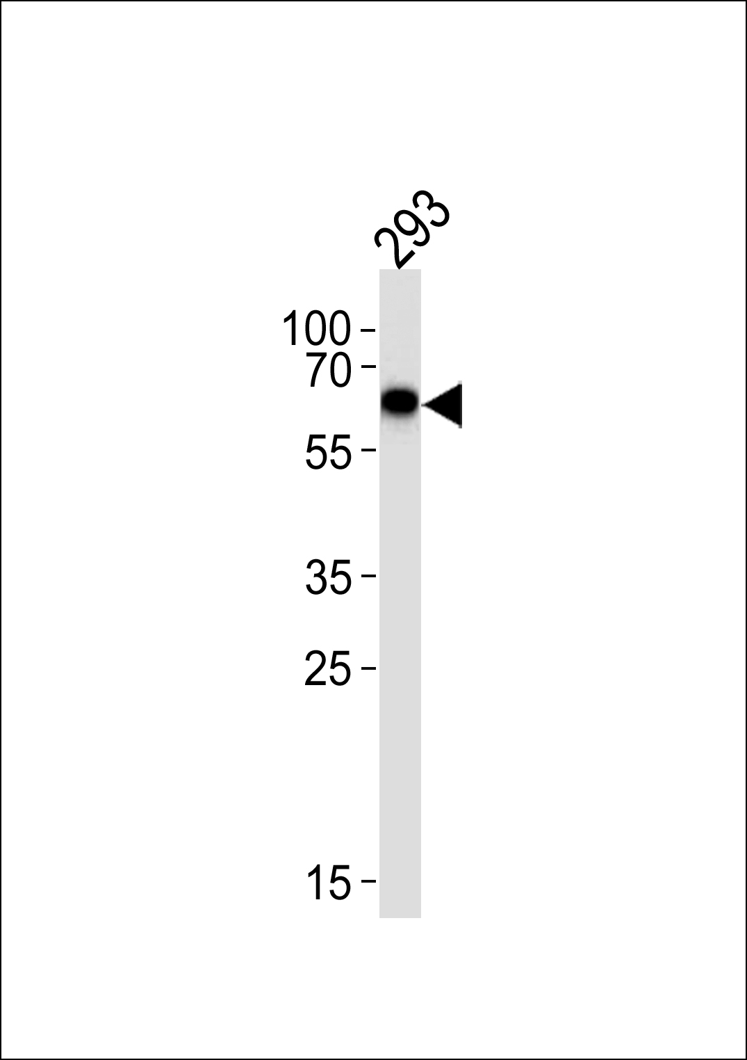
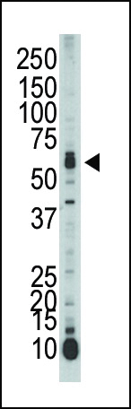
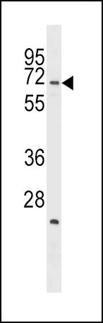
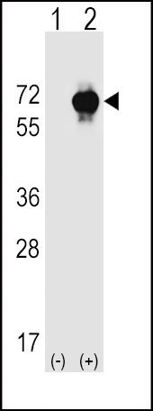
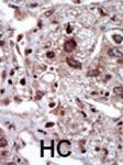
SPAK Antibody (Center)
Purified Rabbit Polyclonal Antibody (Pab)
- 产品详情
- 文献引用 : 4
- 实验流程
- 背景知识
Application
| WB, IHC-P, E |
|---|---|
| Primary Accession | Q9UEW8 |
| Other Accession | O88506, Q9Z1W9 |
| Reactivity | Human, Rat, Mouse |
| Predicted | Rat |
| Host | Rabbit |
| Clonality | Polyclonal |
| Isotype | Rabbit IgG |
| Calculated MW | 59474 Da |
| Antigen Region | 346-376 aa |
| Gene ID | 27347 |
|---|---|
| Other Names | STE20/SPS1-related proline-alanine-rich protein kinase, Ste-20-related kinase, DCHT, Serine/threonine-protein kinase 39, STK39, SPAK |
| Target/Specificity | This SPAK antibody is generated from rabbits immunized with a KLH conjugated synthetic peptide between 346-376 amino acids from the Central region of human SPAK. |
| Dilution | WB~~1:1000 IHC-P~~1:100~500 E~~Use at an assay dependent concentration. |
| Format | Purified polyclonal antibody supplied in PBS with 0.09% (W/V) sodium azide. This antibody is prepared by Saturated Ammonium Sulfate (SAS) precipitation followed by dialysis against PBS. |
| Storage | Maintain refrigerated at 2-8°C for up to 2 weeks. For long term storage store at -20°C in small aliquots to prevent freeze-thaw cycles. |
| Precautions | SPAK Antibody (Center) is for research use only and not for use in diagnostic or therapeutic procedures. |
| Name | STK39 |
|---|---|
| Function | Effector serine/threonine-protein kinase component of the WNK-SPAK/OSR1 kinase cascade, which is involved in various processes, such as ion transport, response to hypertonic stress and blood pressure (PubMed:16669787, PubMed:18270262, PubMed:21321328, PubMed:34289367). Specifically recognizes and binds proteins with a RFXV motif (PubMed:16669787, PubMed:21321328). Acts downstream of WNK kinases (WNK1, WNK2, WNK3 or WNK4): following activation by WNK kinases, catalyzes phosphorylation of ion cotransporters, such as SLC12A1/NKCC2, SLC12A2/NKCC1, SLC12A3/NCC, SLC12A5/KCC2 or SLC12A6/KCC3, regulating their activity (PubMed:21321328). Mediates regulatory volume increase in response to hyperosmotic stress by catalyzing phosphorylation of ion cotransporters SLC12A1/NKCC2, SLC12A2/NKCC1 and SLC12A6/KCC3 downstream of WNK1 and WNK3 kinases (PubMed:12740379, PubMed:16669787, PubMed:21321328). Phosphorylation of Na-K-Cl cotransporters SLC12A2/NKCC1 and SLC12A2/NKCC1 promote their activation and ion influx; simultaneously, phosphorylation of K-Cl cotransporters SLC12A5/KCC2 and SLC12A6/KCC3 inhibit their activity, blocking ion efflux (PubMed:16669787, PubMed:19665974, PubMed:21321328). Acts as a regulator of NaCl reabsorption in the distal nephron by mediating phosphorylation and activation of the thiazide-sensitive Na-Cl cotransporter SLC12A3/NCC in distal convoluted tubule cells of kidney downstream of WNK4 (PubMed:18270262). Mediates the inhibition of SLC4A4, SLC26A6 as well as CFTR activities (By similarity). Phosphorylates RELT (By similarity). |
| Cellular Location | Cytoplasm. Nucleus. Note=Nucleus when caspase-cleaved. |
| Tissue Location | Predominantly expressed in brain and pancreas followed by heart, lung, kidney, skeletal muscle, liver, placenta and testis. |
For Research Use Only. Not For Use In Diagnostic Procedures.

Provided below are standard protocols that you may find useful for product applications.
BACKGROUND
SPAK is a serine/threonine kinase containing an N-terminal series of proline and alanine repeats (PAPA box), followed by a serine/threonine kinase catalytic domain, a nuclear localization signal, a consensus caspase cleavage recognition motif, and a C-terminal region. Northern blot analysis detects ubiquitous expression, most abundantly in brain and pancreas. SPAK can phosphorylate itself and an exogenous substrate in vitro. SPAK immunoprecipitates from transfected mammalian cells in a complex with another serine/threonine kinase that phosphorylates catalytically inactive SPAK. SPAK activates the p38 MAP kinase pathway in cotransfection assays. Full-length SPAK is expressed in the cytoplasm in transfected cells, while a mutant corresponding to caspase-cleaved STK39 localizes predominantly in the nucleus.
REFERENCES
Dowd, B.F., et al., J. Biol. Chem. 278(30):27347-27353 (2003).
Johnston, A.M., et al., Oncogene 19(37):4290-4297 (2000).
终于等到您。ABCEPTA(百远生物)抗体产品。
点击下方“我要评价 ”按钮提交您的反馈信息,您的反馈和评价是我们最宝贵的财富之一,
我们将在1-3个工作日内处理您的反馈信息。
如有疑问,联系:0512-88856768 tech-china@abcepta.com.


