


 癌症的基本特征包括细胞增殖、血管生成、迁移、凋亡逃避机制和细胞永生等。找到癌症发生过程中这些通路的关键标记物和对应的抗体用于检测至关重要。
 为您推荐一个泛素化位点预测神器——泛素化分析工具,可以为您的蛋白的泛素化位点作出预测和评分。
 细胞自噬受体图形绘图工具为你的蛋白的细胞受体结合位点作出预测和评分,识别结合到自噬通路中的蛋白是非常重要的,便于让我们理解自噬在正常生理、病理过程中的作用,如发育、细胞分化、神经退化性疾病、压力条件下、感染和癌症。
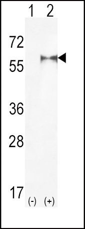
PIP5K1A Antibody (N-term)
Purified Rabbit Polyclonal Antibody (Pab)

- 产品详情
- 文献引用 : 2
- 实验流程
- 背景知识
Application
| WB, E |
|---|---|
| Primary Accession | Q99755 |
| Reactivity | Human |
| Host | Rabbit |
| Clonality | Polyclonal |
| Isotype | Rabbit IgG |
| Calculated MW | 62633 Da |
| Antigen Region | 19-50 aa |
| Gene ID | 8394 |
|---|---|
| Other Names | Phosphatidylinositol 4-phosphate 5-kinase type-1 alpha, PIP5K1-alpha, PtdIns(4)P-5-kinase 1 alpha, 68 kDa type I phosphatidylinositol 4-phosphate 5-kinase alpha, Phosphatidylinositol 4-phosphate 5-kinase type I alpha, PIP5KIalpha, PIP5K1A |
| Target/Specificity | This PIP5K1A antibody is generated from rabbits immunized with a KLH conjugated synthetic peptide between 19-50 amino acids from the N-terminal region of human PIP5K1A. |
| Dilution | WB~~1:1000 E~~Use at an assay dependent concentration. |
| Format | Purified polyclonal antibody supplied in PBS with 0.09% (W/V) sodium azide. This antibody is prepared by Saturated Ammonium Sulfate (SAS) precipitation followed by dialysis against PBS. |
| Storage | Maintain refrigerated at 2-8°C for up to 2 weeks. For long term storage store at -20°C in small aliquots to prevent freeze-thaw cycles. |
| Precautions | PIP5K1A Antibody (N-term) is for research use only and not for use in diagnostic or therapeutic procedures. |
| Name | PIP5K1A (HGNC:8994) |
|---|---|
| Function | Catalyzes the phosphorylation of phosphatidylinositol 4- phosphate (PtdIns(4)P/PI4P) to form phosphatidylinositol 4,5- bisphosphate (PtdIns(4,5)P2/PIP2), a lipid second messenger that regulates several cellular processes such as signal transduction, vesicle trafficking, actin cytoskeleton dynamics, cell adhesion, and cell motility (PubMed:21477596, PubMed:22942276, PubMed:8955136). PtdIns(4,5)P2 can directly act as a second messenger or can be utilized as a precursor to generate other second messengers: inositol 1,4,5- trisphosphate (IP3), diacylglycerol (DAG) or phosphatidylinositol- 3,4,5-trisphosphate (PtdIns(3,4,5)P3/PIP3) (PubMed:19158393, PubMed:20660631). PIP5K1A-mediated phosphorylation of PtdIns(4)P is the predominant pathway for PtdIns(4,5)P2 synthesis (By similarity). Can also use phosphatidylinositol (PtdIns) as substrate in vitro (PubMed:22942276). Together with PIP5K1C, is required for phagocytosis, both enzymes regulating different types of actin remodeling at sequential steps (By similarity). Promotes particle ingestion by activating the WAS GTPase-binding protein that induces Arp2/3 dependent actin polymerization at the nascent phagocytic cup (By similarity). Together with PIP5K1B, is required, after stimulation by G-protein coupled receptors, for the synthesis of IP3 that will induce stable platelet adhesion (By similarity). Recruited to the plasma membrane by the E-cadherin/beta-catenin complex where it provides the substrate PtdIns(4,5)P2 for the production of PtdIns(3,4,5)P3, IP3 and DAG, that will mobilize internal calcium and drive keratinocyte differentiation (PubMed:19158393). Positively regulates insulin-induced translocation of SLC2A4 to the cell membrane in adipocytes (By similarity). Together with PIP5K1C has a role during embryogenesis (By similarity). Independently of its catalytic activity, is required for membrane ruffling formation, actin organization and focal adhesion formation during directional cell migration by controlling integrin-induced translocation of the small GTPase RAC1 to the plasma membrane (PubMed:20660631). Also functions in the nucleus where it acts as an activator of TUT1 adenylyltransferase activity in nuclear speckles, thereby regulating mRNA polyadenylation of a select set of mRNAs (PubMed:18288197). |
| Cellular Location | Cell membrane {ECO:0000250|UniProtKB:P70182}. Cytoplasm {ECO:0000250|UniProtKB:P70182}. Nucleus. Nucleus speckle. Cell projection, ruffle. Cell projection, lamellipodium. Note=Colocalizes with RAC1 at actin-rich membrane ruffles (PubMed:20660631). Localizes to nuclear speckles and associates with TUT1 to regulate polyadenylation of selected mRNAs (PubMed:18288197). |
| Tissue Location | Highly expressed in heart, placenta, skeletal muscle, kidney and pancreas. Detected at lower levels in brain, lung and liver. |
For Research Use Only. Not For Use In Diagnostic Procedures.

Provided below are standard protocols that you may find useful for product applications.
BACKGROUND
Overexpression of phosphatidylinositol phosphate 5-kinase alpha (PIP5KIalpha), which synthesizes PIP2, suppresses apoptosis, whereas a kinase-deficient mutant does not. Protection by the wild-type PIP5KIalpha isaccompanied by decreases in the generation of activated caspases and of caspase 3-cleaved PARP. Protection is not mediated through PIP3 or Akt activation. An anti-apoptotic role for PIP(2) is substantiated by the finding that PIP5KIalpha is cleaved by caspase 3 during apoptosis, and cleavage inactivates PIP5KIalpha in vitro. Mutation of the P(4) position (D279A) of the PIP5KIalpha caspase 3 cleavage consensus prevents cleavage in vitro, and during apoptosis in vivo. Significantly, the caspase 3-resistant PIP5KIalpha mutant is more effective in suppressing apoptosis than the wild-type kinase. PIP2 is a direct regulator of apical and effector caspases in the death receptor and mitochondrial pathways, and PIP5KIalpha inactivation contributes to the progression of apoptosis.
REFERENCES
Doughman, R.L., et al., J. Biol. Chem. 278(25):23036-23045 (2003).
Loijens, J.C., et al., J. Biol. Chem. 271(51):32937-32943 (1996).
Xie, Y., et al., Cytogenet. Cell Genet. 88 (3-4), 197-199 (2000).
终于等到您。ABCEPTA(百远生物)抗体产品。
点击下方“我要评价 ”按钮提交您的反馈信息,您的反馈和评价是我们最宝贵的财富之一,
我们将在1-3个工作日内处理您的反馈信息。
如有疑问,联系:0512-88856768 tech-china@abcepta.com.


