


 癌症的基本特征包括细胞增殖、血管生成、迁移、凋亡逃避机制和细胞永生等。找到癌症发生过程中这些通路的关键标记物和对应的抗体用于检测至关重要。
 为您推荐一个泛素化位点预测神器——泛素化分析工具,可以为您的蛋白的泛素化位点作出预测和评分。
 细胞自噬受体图形绘图工具为你的蛋白的细胞受体结合位点作出预测和评分,识别结合到自噬通路中的蛋白是非常重要的,便于让我们理解自噬在正常生理、病理过程中的作用,如发育、细胞分化、神经退化性疾病、压力条件下、感染和癌症。
MLKLAK Antibody (Center)
Purified Rabbit Polyclonal Antibody (Pab)
- 产品详情
- 文献引用 : 1
- 实验流程
- 背景知识
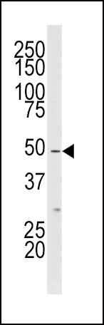
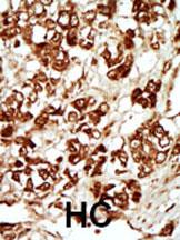
Application
| WB, IHC-P, E |
|---|---|
| Primary Accession | Q9NYL2 |
| Other Accession | Q9ESL4, NP_598407 |
| Reactivity | Human |
| Predicted | Mouse |
| Host | Rabbit |
| Clonality | Polyclonal |
| Isotype | Rabbit IgG |
| Antigen Region | 271-300 aa |
| Other Names | Mitogen-activated protein kinase kinase kinase MLT, Human cervical cancer suppressor gene 4 protein, HCCS-4, Leucine zipper- and sterile alpha motif-containing kinase, MLK-like mitogen-activated protein triple kinase, Mixed lineage kinase-related kinase, MLK-related kinase, MRK, Sterile alpha motif- and leucine zipper-containing kinase AZK, ZAK, MLTK |
|---|---|
| Target/Specificity | This MLKLAK antibody is generated from rabbits immunized with a KLH conjugated synthetic peptide between 271-300 amino acids from the Central region of human MLKLAK. |
| Dilution | WB~~1:1000 IHC-P~~1:100~500 E~~Use at an assay dependent concentration. |
| Format | Purified polyclonal antibody supplied in PBS with 0.09% (W/V) sodium azide. This antibody is prepared by Saturated Ammonium Sulfate (SAS) precipitation followed by dialysis against PBS. |
| Storage | Maintain refrigerated at 2-8°C for up to 2 weeks. For long term storage store at -20°C in small aliquots to prevent freeze-thaw cycles. |
| Precautions | MLKLAK Antibody (Center) is for research use only and not for use in diagnostic or therapeutic procedures. |
For Research Use Only. Not For Use In Diagnostic Procedures.

Provided below are standard protocols that you may find useful for product applications.
BACKGROUND
MLKLAK is a member of the MAPKKK family of signal transduction molecules. It possesses an N-terminal kinase catalytic domain, followed by a leucine zipper motif and a sterile-alpha motif (SAM). This magnesium-binding protein forms homodimers and is located in the cytoplasm. The protein mediates gamma radiation signaling leading to cell cycle arrest and activity of this protein plays a role in cell cycle checkpoint regulation in cells. The protein also has pro-apoptotic activity.
REFERENCES
Blume-Jensen P, et al. Nature 2001. 411: 355.
Cantrell D, J. Cell Sci. 2001. 114: 1439.
Jhiang S Oncogene 2000. 19: 5590.
Manning G, et al. Science 2002. 298: 1912.
Moller, D, et al. Am. J. Physiol. 1994. 266: C351-C359.
Robertson, S. et al. Trends Genet. 2000. 16: 368.
Robinson D, et al. Oncogene 2000. 19: 5548.
Van der Ven, P, et al. Hum. Molec. Genet. 1993. 2: 1889.
Vanhaesebroeck, B, et al. Biochem. J. 2000. 346: 561.
Van Weering D, et al. Recent Results Cancer Res. 1998. 154: 271.
终于等到您。ABCEPTA(百远生物)抗体产品。
点击下方“我要评价 ”按钮提交您的反馈信息,您的反馈和评价是我们最宝贵的财富之一,
我们将在1-3个工作日内处理您的反馈信息。
如有疑问,联系:0512-88856768 tech-china@abcepta.com.


