


 癌症的基本特征包括细胞增殖、血管生成、迁移、凋亡逃避机制和细胞永生等。找到癌症发生过程中这些通路的关键标记物和对应的抗体用于检测至关重要。
 为您推荐一个泛素化位点预测神器——泛素化分析工具,可以为您的蛋白的泛素化位点作出预测和评分。
 细胞自噬受体图形绘图工具为你的蛋白的细胞受体结合位点作出预测和评分,识别结合到自噬通路中的蛋白是非常重要的,便于让我们理解自噬在正常生理、病理过程中的作用,如发育、细胞分化、神经退化性疾病、压力条件下、感染和癌症。
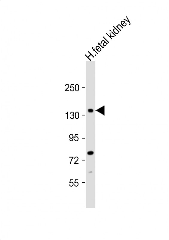
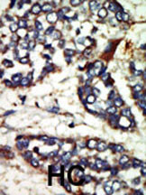
NEK1 Antibody (C-term)
Purified Rabbit Polyclonal Antibody (Pab)
- 产品详情
- 实验流程
- 背景知识
Application
| IHC-P, WB, E |
|---|---|
| Primary Accession | Q96PY6 |
| Other Accession | P51954 |
| Reactivity | Human |
| Predicted | Mouse |
| Host | Rabbit |
| Clonality | Polyclonal |
| Isotype | Rabbit IgG |
| Calculated MW | 142828 Da |
| Antigen Region | 1165-1196 aa |
| Gene ID | 4750 |
|---|---|
| Other Names | Serine/threonine-protein kinase Nek1, Never in mitosis A-related kinase 1, NimA-related protein kinase 1, Renal carcinoma antigen NY-REN-55, NEK1, KIAA1901 |
| Target/Specificity | This NEK1 antibody is generated from rabbits immunized with a KLH conjugated synthetic peptide between 1165-1196 amino acids from the C-terminal region of human NEK1. |
| Dilution | IHC-P~~1:100~500 WB~~1:500 E~~Use at an assay dependent concentration. |
| Format | Purified polyclonal antibody supplied in PBS with 0.09% (W/V) sodium azide. This antibody is prepared by Saturated Ammonium Sulfate (SAS) precipitation followed by dialysis against PBS. |
| Storage | Maintain refrigerated at 2-8°C for up to 2 weeks. For long term storage store at -20°C in small aliquots to prevent freeze-thaw cycles. |
| Precautions | NEK1 Antibody (C-term) is for research use only and not for use in diagnostic or therapeutic procedures. |
| Name | NEK1 |
|---|---|
| Synonyms | KIAA1901 |
| Function | Phosphorylates serines and threonines, but also appears to possess tyrosine kinase activity (PubMed:20230784). Involved in DNA damage checkpoint control and for proper DNA damage repair (PubMed:20230784). In response to injury that includes DNA damage, NEK1 phosphorylates VDAC1 to limit mitochondrial cell death (PubMed:20230784). May be implicated in the control of meiosis (By similarity). Involved in cilium assembly (PubMed:21211617). |
| Cellular Location | Nucleus. Cytoplasm, cytoskeleton, microtubule organizing center, centrosome {ECO:0000250|UniProtKB:P51954}. Cytoplasm. Note=Associated with the pericentriolar material (PubMed:21211617). Localizes to centrosome during interphase and mitosis (By similarity). Translocated from cytoplasm to discrete nuclear foci at sites of DNA damage (PubMed:15604234) {ECO:0000250|UniProtKB:P51954, ECO:0000269|PubMed:15604234, ECO:0000269|PubMed:21211617} |
| Tissue Location | High fetal expression in the brain and kidney. |
For Research Use Only. Not For Use In Diagnostic Procedures.
Provided below are standard protocols that you may find useful for product applications.
BACKGROUND
Protein kinases are enzymes that transfer a phosphate group from a phosphate donor, generally the g phosphate of ATP, onto an acceptor amino acid in a substrate protein. By this basic mechanism, protein kinases mediate most of the signal transduction in eukaryotic cells, regulating cellular metabolism, transcription, cell cycle progression, cytoskeletal rearrangement and cell movement, apoptosis, and differentiation. With more than 500 gene products, the protein kinase family is one of the largest families of proteins in eukaryotes. The family has been classified in 8 major groups based on sequence comparison of their tyrosine (PTK) or serine/threonine (STK) kinase catalytic domains. The STE group (homologs of yeast Sterile 7, 11, 20 kinases) consists of 50 kinases related to the mitogen-activated protein kinase (MAPK) cascade families (Ste7/MAP2K, Ste11/MAP3K, and Ste20/MAP4K). MAP kinase cascades, consisting of a MAPK and one or more upstream regulatory kinases (MAPKKs) have been best characterized in the yeast pheromone response pathway. Pheromones bind to Ste cell surface receptors and activate yeast MAPK pathway.
REFERENCES
Surpili, M.J., et al., Biochemistry 42(51):15369-15376 (2003).
Scanlan, M.J., et al., Int. J. Cancer 83(4):456-464 (1999).
Letwin, K., et al., EMBO J. 11(10):3521-3531 (1992).
终于等到您。ABCEPTA(百远生物)抗体产品。
点击下方“我要评价 ”按钮提交您的反馈信息,您的反馈和评价是我们最宝贵的财富之一,
我们将在1-3个工作日内处理您的反馈信息。
如有疑问,联系:0512-88856768 tech-china@abcepta.com.


