


 癌症的基本特征包括细胞增殖、血管生成、迁移、凋亡逃避机制和细胞永生等。找到癌症发生过程中这些通路的关键标记物和对应的抗体用于检测至关重要。
 为您推荐一个泛素化位点预测神器——泛素化分析工具,可以为您的蛋白的泛素化位点作出预测和评分。
 细胞自噬受体图形绘图工具为你的蛋白的细胞受体结合位点作出预测和评分,识别结合到自噬通路中的蛋白是非常重要的,便于让我们理解自噬在正常生理、病理过程中的作用,如发育、细胞分化、神经退化性疾病、压力条件下、感染和癌症。
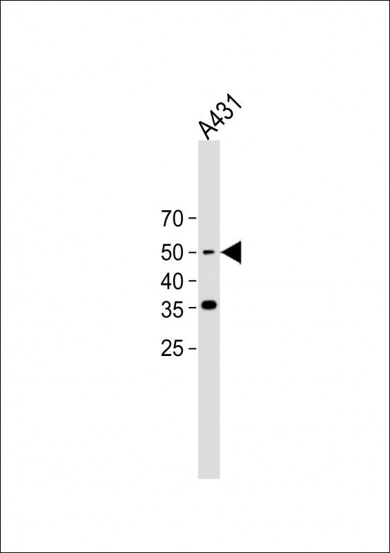
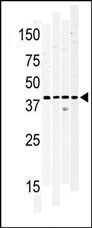
WNK1 Antibody (Center)
Purified Rabbit Polyclonal Antibody (Pab)
- 产品详情
- 实验流程
- 背景知识
Application
| WB, E |
|---|---|
| Primary Accession | Q9H4A3 |
| Other Accession | Q9JIH7, P83741, Q4VBX9 |
| Reactivity | Human, Mouse |
| Predicted | Rat |
| Host | Rabbit |
| Clonality | Polyclonal |
| Isotype | Rabbit IgG |
| Calculated MW | 250794 Da |
| Gene ID | 65125 |
|---|---|
| Other Names | Serine/threonine-protein kinase WNK1, Erythrocyte 65 kDa protein, p65, Kinase deficient protein, Protein kinase lysine-deficient 1, Protein kinase with no lysine 1, hWNK1, WNK1, HSN2, KDP, KIAA0344, PRKWNK1 |
| Target/Specificity | This WNK1 antibody is generated from rabbits immunized with a KLH conjugated synthetic peptide selected from the center region of human WNK1. |
| Dilution | WB~~1:1000 E~~Use at an assay dependent concentration. |
| Format | Purified polyclonal antibody supplied in PBS with 0.05% (V/V) Proclin 300. This antibody is purified through a protein A column, followed by peptide affinity purification. |
| Storage | Maintain refrigerated at 2-8°C for up to 2 weeks. For long term storage store at -20°C in small aliquots to prevent freeze-thaw cycles. |
| Precautions | WNK1 Antibody (Center) is for research use only and not for use in diagnostic or therapeutic procedures. |
| Name | WNK1 {ECO:0000303|PubMed:11571656, ECO:0000312|HGNC:HGNC:14540} |
|---|---|
| Function | Serine/threonine-protein kinase component of the WNK1- SPAK/OSR1 kinase cascade, which acts as a key regulator of blood pressure and regulatory volume increase by promoting ion influx (PubMed:15883153, PubMed:17190791, PubMed:31656913, PubMed:34289367, PubMed:36318922). WNK1 mediates regulatory volume increase in response to hyperosmotic stress by acting as a molecular crowding sensor, which senses cell shrinkage and mediates formation of a membraneless compartment by undergoing liquid-liquid phase separation (PubMed:36318922). The membraneless compartment concentrates WNK1 with its substrates, OXSR1/OSR1 and STK39/SPAK, promoting WNK1-dependent phosphorylation and activation of downstream kinases OXSR1/OSR1 and STK39/SPAK (PubMed:15883153, PubMed:16263722, PubMed:17190791, PubMed:19739668, PubMed:21321328, PubMed:22989884, PubMed:25477473, PubMed:34289367, PubMed:36318922). Following activation, OXSR1/OSR1 and STK39/SPAK catalyze phosphorylation of ion cotransporters SLC12A1/NKCC2, SLC12A2/NKCC1, SLC12A5/KCC2 and SLC12A6/KCC3, regulating their activity (PubMed:16263722, PubMed:21321328). Phosphorylation of Na-K-Cl cotransporters SLC12A2/NKCC1 and SLC12A2/NKCC1 promote their activation and ion influx; simultaneously, phosphorylation of K-Cl cotransporters SLC12A5/KCC2 and SLC12A6/KCC3 inhibit their activity, blocking ion efflux (PubMed:19665974, PubMed:21321328). Also acts as a regulator of angiogenesis in endothelial cells via activation of OXSR1/OSR1 and STK39/SPAK: activation of OXSR1/OSR1 regulates chemotaxis and invasion, while STK39/SPAK regulates endothelial cell proliferation (PubMed:25362046). Also acts independently of the WNK1- SPAK/OSR1 kinase cascade by catalyzing phosphorylation of other substrates, such as SYT2, PCF11 and NEDD4L (PubMed:29196535). Mediates phosphorylation of SYT2, regulating SYT2 association with phospholipids and membrane-binding (By similarity). Regulates mRNA export in the nucleus by mediating phosphorylation of PCF11, thereby decreasing the association between PCF11 and POLR2A/RNA polymerase II and promoting mRNA export to the cytoplasm (PubMed:29196535). Acts as a negative regulator of autophagy (PubMed:27911840). Required for the abscission step during mitosis, independently of the WNK1-SPAK/OSR1 kinase cascade (PubMed:21220314). May also play a role in actin cytoskeletal reorganization (PubMed:10660600). Also acts as a scaffold protein independently of its protein kinase activity: negatively regulates cell membrane localization of various transporters and channels, such as SLC4A4, SLC26A6, SLC26A9, TRPV4 and CFTR (By similarity). Involved in the regulation of epithelial Na(+) channel (ENaC) by promoting activation of SGK1 in a kinase-independent manner: probably acts as a scaffold protein that promotes the recruitment of SGK1 to the mTORC2 complex in response to chloride, leading to mTORC2-dependent phosphorylation and activation of SGK1 (PubMed:36373794). Acts as an assembly factor for the ER membrane protein complex independently of its protein kinase activity: associates with EMC2 in the cytoplasm via its amphipathic alpha-helix, and prevents EMC2 ubiquitination and subsequent degradation, thereby promoting EMC2 stabilization (PubMed:33964204). |
| Cellular Location | Cytoplasm. Nucleus. Cytoplasm, cytoskeleton, spindle. Note=Mediates formation and localizes to cytoplasmic membraneless compartment in response to hyperosmotic stress (PubMed:36318922). Also localizes to the nucleus (PubMed:29196535) Localizes to the mitotic spindle during mitosis (PubMed:21220314) |
| Tissue Location | Widely expressed, with highest levels observed in the testis, heart, kidney and skeletal muscle [Isoform 3]: This isoform is kidney-specific and specifically expressed in the distal convoluted tubule (DCT) and connecting tubule (CNT) of the nephron. |
Research Areas
For Research Use Only. Not For Use In Diagnostic Procedures.
Application Protocols
Provided below are standard protocols that you may find useful for product applications.
BACKGROUND
The WNK1 gene encodes a cytoplasmic serine-threonine kinase expressed in distal nephron.[supplied by OMIM]
REFERENCES
Xu, B.E., et al., J. Biol. Chem. 277(50):48456-48462 (2002).
Verissimo, F., et al., Oncogene 20(39):5562-5569 (2001).
Wilson, F.H., et al., Science 293(5532):1107-1112 (2001).
Moore, T.M., et al., J. Biol. Chem. 275(6):4311-4322 (2000).
终于等到您。ABCEPTA(百远生物)抗体产品。
点击下方“我要评价 ”按钮提交您的反馈信息,您的反馈和评价是我们最宝贵的财富之一,
我们将在1-3个工作日内处理您的反馈信息。
如有疑问,联系:0512-88856768 tech-china@abcepta.com.
¥ 1,250.00
Cat# AP8107c


