


 癌症的基本特征包括细胞增殖、血管生成、迁移、凋亡逃避机制和细胞永生等。找到癌症发生过程中这些通路的关键标记物和对应的抗体用于检测至关重要。
 为您推荐一个泛素化位点预测神器——泛素化分析工具,可以为您的蛋白的泛素化位点作出预测和评分。
 细胞自噬受体图形绘图工具为你的蛋白的细胞受体结合位点作出预测和评分,识别结合到自噬通路中的蛋白是非常重要的,便于让我们理解自噬在正常生理、病理过程中的作用,如发育、细胞分化、神经退化性疾病、压力条件下、感染和癌症。
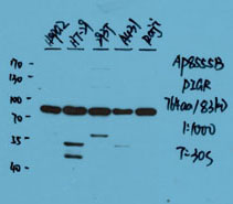
PIGR Antibody (C-term)
Affinity Purified Rabbit Polyclonal Antibody (Pab)
- 产品详情
- 实验流程
- 背景知识
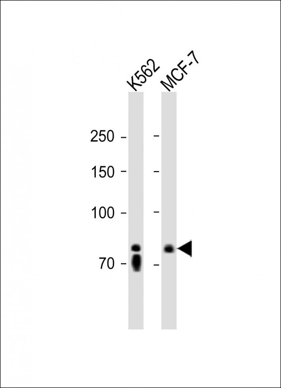
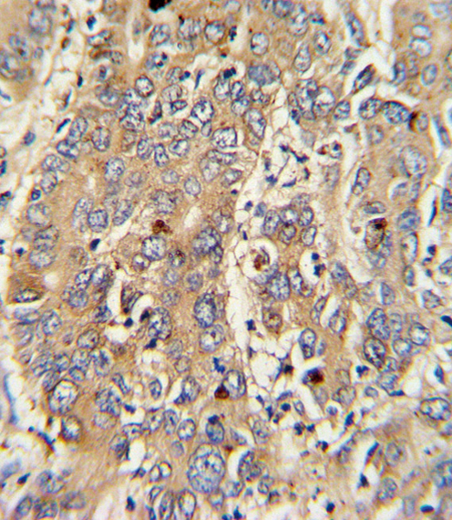
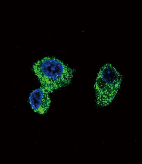
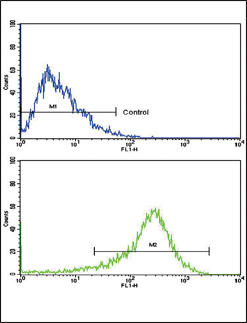
Application
| WB, IHC-P, IF, FC, E |
|---|---|
| Primary Accession | P01833 |
| Reactivity | Human |
| Host | Rabbit |
| Clonality | Polyclonal |
| Isotype | Rabbit IgG |
| Calculated MW | 83284 Da |
| Antigen Region | 646-672 aa |
| Gene ID | 5284 |
|---|---|
| Other Names | Polymeric immunoglobulin receptor, PIgR, Poly-Ig receptor, Hepatocellular carcinoma-associated protein TB6, Secretory component, PIGR |
| Target/Specificity | This PIGR antibody is generated from rabbits immunized with a KLH conjugated synthetic peptide between 646-672 amino acids from the C-terminal region of human PIGR. |
| Dilution | WB~~1:1000 IHC-P~~1:100~500 IF~~1:10~50 FC~~1:10~50 E~~Use at an assay dependent concentration. |
| Format | Purified polyclonal antibody supplied in PBS with 0.05% (V/V) Proclin 300. This antibody is purified through a protein A column, followed by peptide affinity purification. |
| Storage | Maintain refrigerated at 2-8°C for up to 2 weeks. For long term storage store at -20°C in small aliquots to prevent freeze-thaw cycles. |
| Precautions | PIGR Antibody (C-term) is for research use only and not for use in diagnostic or therapeutic procedures. |
| Name | PIGR |
|---|---|
| Function | [Polymeric immunoglobulin receptor]: Mediates selective transcytosis of polymeric IgA and IgM across mucosal epithelial cells. Binds polymeric IgA and IgM at the basolateral surface of epithelial cells. The complex is then transported across the cell to be secreted at the apical surface. During this process, a cleavage occurs that separates the extracellular (known as the secretory component) from the transmembrane segment. |
| Cellular Location | [Polymeric immunoglobulin receptor]: Cell membrane; Single-pass type I membrane protein |
For Research Use Only. Not For Use In Diagnostic Procedures.
Provided below are standard protocols that you may find useful for product applications.
BACKGROUND
PIGR binds polymeric IgA and IgM at the basolateral surface of epithelial cells. The complex is then transported across the cell to be secreted at the apical surface. During this process a cleavage occurs that separates the extracellular (known as the secretory component) from the transmembrane segment.
REFERENCES
Ewing,R.M., et.al., Mol. Syst. Biol. 3, 89 (2007)
Orzech,E., Cohen,S., et.al., J. Biol. Chem. 275 (20), 15207-15219 (2000)
终于等到您。ABCEPTA(百远生物)抗体产品。
点击下方“我要评价 ”按钮提交您的反馈信息,您的反馈和评价是我们最宝贵的财富之一,
我们将在1-3个工作日内处理您的反馈信息。
如有疑问,联系:0512-88856768 tech-china@abcepta.com.
|
Anonymous
2016-08-24 09:20:44
1
2
3
4
5
|
Species tested
Human
Application tested
WB
Organization tested
HepG2,HT-29,293T,A431,Raji
Barcode encoding
Brief protocol
1. Block with 3% skim milk for 1 hour at room temperature.
2. Incubate overnight with Abgent primary antibody 1:1000 in 3% skim milk at 4℃
3. Wash 5*5 min with TBST.
4. Incubate with HRP-conjugated secondary antibody 1:5000 in 3% skim milk for 1 hour at room temperature.
5. Wash 5*5 min with TBST.
6. Incubate with ECL substrates and expose
|
|


