


 癌症的基本特征包括细胞增殖、血管生成、迁移、凋亡逃避机制和细胞永生等。找到癌症发生过程中这些通路的关键标记物和对应的抗体用于检测至关重要。
 为您推荐一个泛素化位点预测神器——泛素化分析工具,可以为您的蛋白的泛素化位点作出预测和评分。
 细胞自噬受体图形绘图工具为你的蛋白的细胞受体结合位点作出预测和评分,识别结合到自噬通路中的蛋白是非常重要的,便于让我们理解自噬在正常生理、病理过程中的作用,如发育、细胞分化、神经退化性疾病、压力条件下、感染和癌症。
CLIP1 Antibody (N-term)
Affinity Purified Rabbit Polyclonal Antibody (Pab)
- 产品详情
- 实验流程
- 背景知识
Application
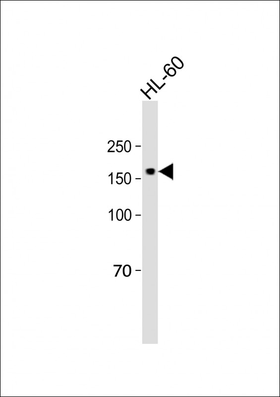
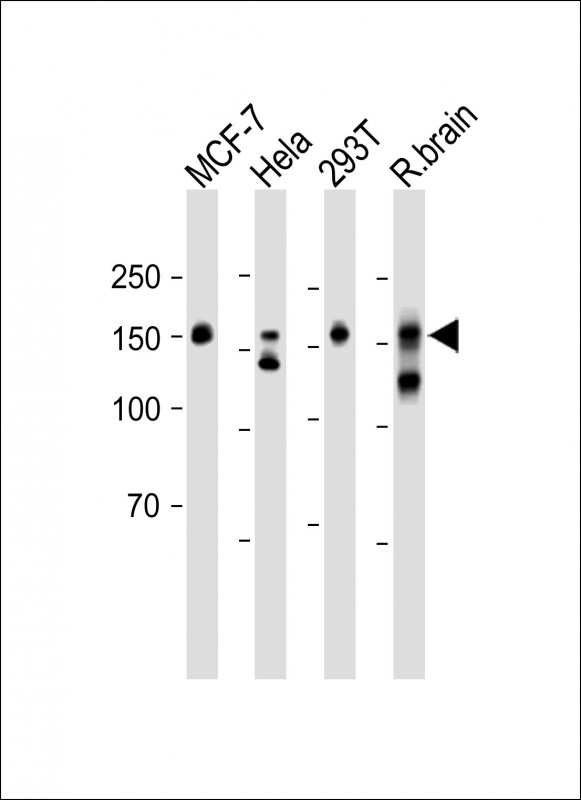
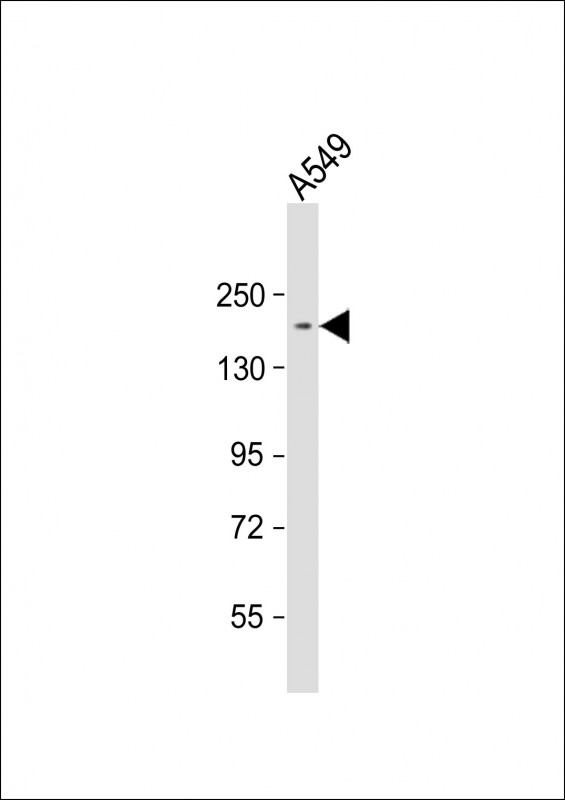
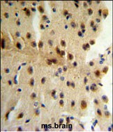
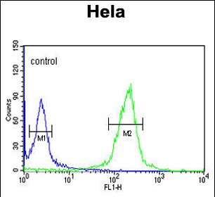
| WB, IHC-P, FC, E |
|---|---|
| Primary Accession | P30622 |
| Other Accession | O55156, Q9Z0H8, Q9UDT6, Q922J3, O42184 |
| Reactivity | Human |
| Predicted | Chicken, Rat |
| Host | Rabbit |
| Clonality | Polyclonal |
| Isotype | Rabbit IgG |
| Calculated MW | 162246 Da |
| Antigen Region | 228-254 aa |
| Gene ID | 6249 |
|---|---|
| Other Names | CAP-Gly domain-containing linker protein 1, Cytoplasmic linker protein 1, Cytoplasmic linker protein 170 alpha-2, CLIP-170, Reed-Sternberg intermediate filament-associated protein, Restin, CLIP1, CYLN1, RSN |
| Target/Specificity | This CLIP1 antibody is generated from rabbits immunized with a KLH conjugated synthetic peptide between 228-254 amino acids from the N-terminal region of human CLIP1. |
| Dilution | WB~~1:1000 IHC-P~~1:100~500 FC~~1:10~50 E~~Use at an assay dependent concentration. |
| Format | Purified polyclonal antibody supplied in PBS with 0.09% (W/V) sodium azide. This antibody is purified through a protein A column, followed by peptide affinity purification. |
| Storage | Maintain refrigerated at 2-8°C for up to 2 weeks. For long term storage store at -20°C in small aliquots to prevent freeze-thaw cycles. |
| Precautions | CLIP1 Antibody (N-term) is for research use only and not for use in diagnostic or therapeutic procedures. |
| Name | CLIP1 |
|---|---|
| Synonyms | CYLN1, RSN |
| Function | Binds to the plus end of microtubules and regulates the dynamics of the microtubule cytoskeleton. Promotes microtubule growth and microtubule bundling. Links cytoplasmic vesicles to microtubules and thereby plays an important role in intracellular vesicle trafficking. Plays a role macropinocytosis and endosome trafficking. |
| Cellular Location | Cytoplasm. Cytoplasm, cytoskeleton. Cytoplasmic vesicle membrane; Peripheral membrane protein; Cytoplasmic side. Cell projection, ruffle. Note=Localizes to microtubule plus ends (PubMed:17889670, PubMed:21646404). Localizes preferentially to the ends of tyrosinated microtubules (By similarity). Accumulates in plasma membrane regions with ruffling and protrusions. Associates with the membranes of intermediate macropinocytic vesicles (PubMed:12433698) {ECO:0000250|UniProtKB:Q922J3, ECO:0000269|PubMed:12433698, ECO:0000269|PubMed:17889670, ECO:0000269|PubMed:21646404} |
| Tissue Location | Detected in dendritic cells (at protein level). Highly expressed in the Reed-Sternberg cells of Hodgkin disease |
Research Areas
For Research Use Only. Not For Use In Diagnostic Procedures.
Application Protocols
Provided below are standard protocols that you may find useful for product applications.
BACKGROUND
CLIP1 may be a intermediate filament associated protein that links endocytic vesicles to microtubules.
REFERENCES
Yang,X., et.al., J. Biol. Chem. 284 (42), 28775-28782 (2009)
Meunier,B., et.al., Eur. J. Cell Biol. 88 (2), 91-102 (2009)
终于等到您。ABCEPTA(百远生物)抗体产品。
点击下方“我要评价 ”按钮提交您的反馈信息,您的反馈和评价是我们最宝贵的财富之一,
我们将在1-3个工作日内处理您的反馈信息。
如有疑问,联系:0512-88856768 tech-china@abcepta.com.
¥ 1,250.00
Cat# AP8950a


