


 癌症的基本特征包括细胞增殖、血管生成、迁移、凋亡逃避机制和细胞永生等。找到癌症发生过程中这些通路的关键标记物和对应的抗体用于检测至关重要。
 为您推荐一个泛素化位点预测神器——泛素化分析工具,可以为您的蛋白的泛素化位点作出预测和评分。
 细胞自噬受体图形绘图工具为你的蛋白的细胞受体结合位点作出预测和评分,识别结合到自噬通路中的蛋白是非常重要的,便于让我们理解自噬在正常生理、病理过程中的作用,如发育、细胞分化、神经退化性疾病、压力条件下、感染和癌症。
PRC1 Antibody
Rabbit mAb
- 产品详情
- 实验流程
Application
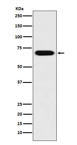
| WB, IHC, IF, FC, ICC, IP, IHF |
|---|---|
| Primary Accession | O43663 |
| Reactivity | Rat, Human, Mouse |
| Clonality | Monoclonal |
| Other Names | ASE1; PRC1; Protein regulating cytokinesis 1; |
| Isotype | Rabbit IgG |
| Host | Rabbit |
| Calculated MW | 71607 Da |
| Dilution | WB 1:500~1:2000 IHC 1:50~1:200 ICC/IF 1:50~1:200 IP 1:50 FC 1:50 |
|---|---|
| Purification | Affinity-chromatography |
| Immunogen | A synthesized peptide derived from human PRC1 |
| Description | Cross-links antiparrallel microtubules at an average distance of 35 nM. Essential for controlling the spatiotemporal formation of the midzone and successful cytokinesis. Required for KIF14 localization to the central spindle and midbody. |
| Storage Condition and Buffer | Rabbit IgG in phosphate buffered saline , pH 7.4, 150mM NaCl, 0.02% sodium azide and 50% glycerol. Store at +4°C short term. Store at -20°C long term. Avoid freeze / thaw cycle. |
| Name | PRC1 (HGNC:9341) |
|---|---|
| Function | Key regulator of cytokinesis that cross-links antiparrallel microtubules at an average distance of 35 nM. Essential for controlling the spatiotemporal formation of the midzone and successful cytokinesis. Required for KIF14 localization to the central spindle and midbody. Required to recruit PLK1 to the spindle. Stimulates PLK1 phosphorylation of RACGAP1 to allow recruitment of ECT2 to the central spindle. Acts as an oncogene for promoting bladder cancer cells proliferation, apoptosis inhibition and carcinogenic progression (PubMed:17409436). |
| Cellular Location | Nucleus. Cytoplasm. Cytoplasm, cytoskeleton, spindle pole. Midbody. Chromosome. Note=Colocalized with KIF20B in the nucleus of bladder carcinoma cells at the interphase. Colocalized with KIF20B in bladder carcinoma cells at prophase, metaphase, early anaphase, at the midzone in late anaphase and at the contractile ring in telophase (PubMed:17409436). Predominantly localized to the nucleus of interphase cells. During mitosis becomes associated with the mitotic spindle poles and localizes with the cell midbody during cytokinesis Co-localizes with PRC1 in early mitosis and at the spindle midzone from anaphase B to telophase (PubMed:15297875, PubMed:15625105) |
| Tissue Location | Overexpressed in bladder cancer cells (PubMed:17409436). |
Research Areas
For Research Use Only. Not For Use In Diagnostic Procedures.
Application Protocols
Provided below are standard protocols that you may find useful for product applications.
终于等到您。ABCEPTA(百远生物)抗体产品。
点击下方“我要评价 ”按钮提交您的反馈信息,您的反馈和评价是我们最宝贵的财富之一,
我们将在1-3个工作日内处理您的反馈信息。
如有疑问,联系:0512-88856768 tech-china@abcepta.com.
¥ 1,500.00
Cat# AP90071


