


 癌症的基本特征包括细胞增殖、血管生成、迁移、凋亡逃避机制和细胞永生等。找到癌症发生过程中这些通路的关键标记物和对应的抗体用于检测至关重要。
 为您推荐一个泛素化位点预测神器——泛素化分析工具,可以为您的蛋白的泛素化位点作出预测和评分。
 细胞自噬受体图形绘图工具为你的蛋白的细胞受体结合位点作出预测和评分,识别结合到自噬通路中的蛋白是非常重要的,便于让我们理解自噬在正常生理、病理过程中的作用,如发育、细胞分化、神经退化性疾病、压力条件下、感染和癌症。
JIP1 Antibody (C-term)
Affinity Purified Rabbit Polyclonal Antibody (Pab)
- 产品详情
- 实验流程
- 背景知识
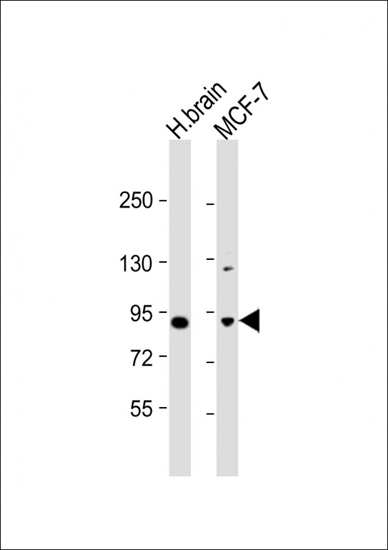
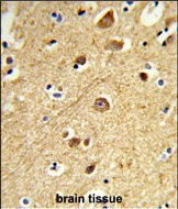
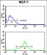
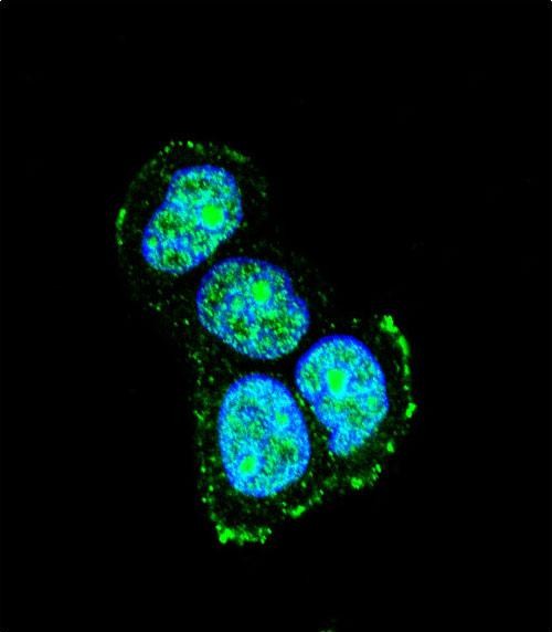
Application
| IHC-P, FC, IF, WB, E |
|---|---|
| Primary Accession | Q9UQF2 |
| Reactivity | Human |
| Host | Rabbit |
| Clonality | Polyclonal |
| Isotype | Rabbit IgG |
| Calculated MW | 77524 Da |
| Antigen Region | 470-498 aa |
| Gene ID | 9479 |
|---|---|
| Other Names | C-Jun-amino-terminal kinase-interacting protein 1, JIP-1, JNK-interacting protein 1, Islet-brain 1, IB-1, JNK MAP kinase scaffold protein 1, Mitogen-activated protein kinase 8-interacting protein 1, MAPK8IP1, IB1, JIP1, PRKM8IP |
| Target/Specificity | This JIP1 antibody is generated from rabbits immunized with a KLH conjugated synthetic peptide between 470-498 amino acids from the C-terminal region of human JIP1. |
| Dilution | IHC-P~~1:100~500 FC~~1:10~50 IF~~1:10~50 WB~~1:1000 E~~Use at an assay dependent concentration. |
| Format | Purified polyclonal antibody supplied in PBS with 0.09% (W/V) sodium azide. This antibody is purified through a protein A column, followed by peptide affinity purification. |
| Storage | Maintain refrigerated at 2-8°C for up to 2 weeks. For long term storage store at -20°C in small aliquots to prevent freeze-thaw cycles. |
| Precautions | JIP1 Antibody (C-term) is for research use only and not for use in diagnostic or therapeutic procedures. |
| Name | MAPK8IP1 |
|---|---|
| Synonyms | IB1, JIP1, PRKM8IP |
| Function | The JNK-interacting protein (JIP) group of scaffold proteins selectively mediates JNK signaling by aggregating specific components of the MAPK cascade to form a functional JNK signaling module. Required for JNK activation in response to excitotoxic stress. Cytoplasmic MAPK8IP1 causes inhibition of JNK-regulated activity by retaining JNK in the cytoplasm and inhibiting JNK phosphorylation of c-Jun. May also participate in ApoER2-specific reelin signaling. Directly, or indirectly, regulates GLUT2 gene expression and beta-cell function. Appears to have a role in cell signaling in mature and developing nerve terminals. May function as a regulator of vesicle transport, through interactions with the JNK-signaling components and motor proteins. Functions as an anti-apoptotic protein and whose level seems to influence the beta-cell death or survival response. Acts as a scaffold protein that coordinates with SH3RF1 in organizing different components of the JNK pathway, including RAC1 or RAC2, MAP3K11/MLK3 or MAP3K7/TAK1, MAP2K7/MKK7, MAPK8/JNK1 and/or MAPK9/JNK2 into a functional multiprotein complex to ensure the effective activation of the JNK signaling pathway. Regulates the activation of MAPK8/JNK1 and differentiation of CD8(+) T-cells. |
| Cellular Location | Cytoplasm. Cytoplasm, perinuclear region. Nucleus. Endoplasmic reticulum membrane. Mitochondrion membrane. Note=Accumulates in cell surface projections. Under certain stress conditions, translocates to the perinuclear region of neurons. In insulin-secreting cells, detected in both the cytoplasm and nucleus (By similarity). |
| Tissue Location | Highly expressed in brain. Expressed in neurons, localizing to neurite tips in differentiating cells. Also expressed in the pancreas, testis and prostate. Low levels in heart, ovary and small intestine. Decreased levels in pancreatic beta cells sensitize cells to IL-1-beta-induced apoptosis |
For Research Use Only. Not For Use In Diagnostic Procedures.
Provided below are standard protocols that you may find useful for product applications.
BACKGROUND
JIP1 is a regulator of the pancreatic beta-cell function. It is highly similar to JIP-1, a mouse protein known to be a regulator of c-Jun amino-terminal kinase (Mapk8). This protein has been shown to prevent MAPK8 mediated activation of transcription factors, and decrease IL-1 beta and MAP kinase kinase 1 (MEKK1) induced apoptosis in pancreatic beta cells. This protein also functions as a DNA-binding transactivator of the glucose transporter GLUT2.
REFERENCES
Mooser,V., et.al., Genomics 55 (2), 202-208 (1999)
终于等到您。ABCEPTA(百远生物)抗体产品。
点击下方“我要评价 ”按钮提交您的反馈信息,您的反馈和评价是我们最宝贵的财富之一,
我们将在1-3个工作日内处理您的反馈信息。
如有疑问,联系:0512-88856768 tech-china@abcepta.com.


