


 癌症的基本特征包括细胞增殖、血管生成、迁移、凋亡逃避机制和细胞永生等。找到癌症发生过程中这些通路的关键标记物和对应的抗体用于检测至关重要。
 为您推荐一个泛素化位点预测神器——泛素化分析工具,可以为您的蛋白的泛素化位点作出预测和评分。
 细胞自噬受体图形绘图工具为你的蛋白的细胞受体结合位点作出预测和评分,识别结合到自噬通路中的蛋白是非常重要的,便于让我们理解自噬在正常生理、病理过程中的作用,如发育、细胞分化、神经退化性疾病、压力条件下、感染和癌症。
Phospho-Smad3 (S423 + S425) Antibody
Rabbit mAb
- 产品详情
- 实验流程
Application
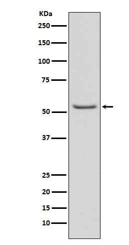
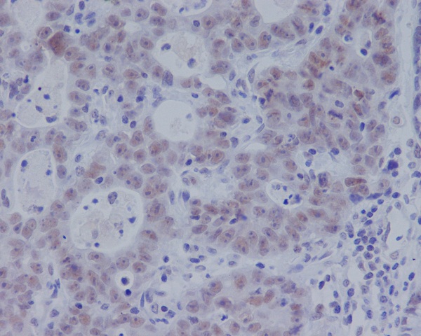
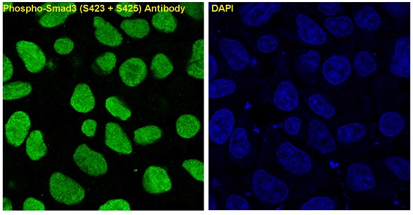
| WB, IHC, IF, ICC, IHF |
|---|---|
| Primary Accession | P84022 |
| Reactivity | Human, Mouse |
| Clonality | Monoclonal |
| Other Names | JV15-2, MAD-3, MADH3, Mad3, Mothers against DPP homolog 3, Mothers against decapentaplegic homolog 3, SMAD 3, Smad 3 |
| Isotype | Rabbit IgG |
| Host | Rabbit |
| Calculated MW | 48081 Da |
| Dilution | WB 1:500~1:2000 IHC 1:50~1:200 ICC/IF 1:50~1:200 |
|---|---|
| Purification | Affinity-chromatography |
| Immunogen | A synthesized peptide derived from human Phospho-Smad3 (S423 + S425) |
| Description | Receptor-regulated SMAD (R-SMAD) that is an intracellular signal transducer and transcriptional modulator activated by TGF-beta (transforming growth factor) and activin type 1 receptor kinases. Binds the TRE element in the promoter region of many genes that are regulated by TGF-beta and, on formation of the SMAD3/SMAD4 complex, activates transcription. |
| Storage Condition and Buffer | Rabbit IgG in phosphate buffered saline , pH 7.4, 150mM NaCl, 0.02% sodium azide and 50% glycerol. Store at +4°C short term. Store at -20°C long term. Avoid freeze / thaw cycle. |
| Name | SMAD3 |
|---|---|
| Synonyms | MADH3 |
| Function | Receptor-regulated SMAD (R-SMAD) that is an intracellular signal transducer and transcriptional modulator activated by TGF-beta (transforming growth factor) and activin type 1 receptor kinases. Binds the TRE element in the promoter region of many genes that are regulated by TGF-beta and, on formation of the SMAD3/SMAD4 complex, activates transcription. Also can form a SMAD3/SMAD4/JUN/FOS complex at the AP- 1/SMAD site to regulate TGF-beta-mediated transcription. Has an inhibitory effect on wound healing probably by modulating both growth and migration of primary keratinocytes and by altering the TGF-mediated chemotaxis of monocytes. This effect on wound healing appears to be hormone-sensitive. Regulator of chondrogenesis and osteogenesis and inhibits early healing of bone fractures. Positively regulates PDPK1 kinase activity by stimulating its dissociation from the 14-3-3 protein YWHAQ which acts as a negative regulator. |
| Cellular Location | Cytoplasm. Nucleus. Note=Cytoplasmic and nuclear in the absence of TGF-beta. On TGF-beta stimulation, migrates to the nucleus when complexed with SMAD4 (PubMed:15799969, PubMed:21145499). Through the action of the phosphatase PPM1A, released from the SMAD2/SMAD4 complex, and exported out of the nucleus by interaction with RANBP1 (PubMed:16751101, PubMed:19289081). Co-localizes with LEMD3 at the nucleus inner membrane (PubMed:15601644). MAPK-mediated phosphorylation appears to have no effect on nuclear import (PubMed:19218245). PDPK1 prevents its nuclear translocation in response to TGF-beta (PubMed:17327236). Localized mainly to the nucleus in the early stages of embryo development with expression becoming evident in the cytoplasm of the inner cell mass at the blastocyst stage (By similarity) {ECO:0000250|UniProtKB:Q8BUN5, ECO:0000269|PubMed:15601644, ECO:0000269|PubMed:15799969, ECO:0000269|PubMed:16751101, ECO:0000269|PubMed:17327236, ECO:0000269|PubMed:19218245, ECO:0000269|PubMed:19289081, ECO:0000269|PubMed:21145499} |
Research Areas
For Research Use Only. Not For Use In Diagnostic Procedures.
Application Protocols
Provided below are standard protocols that you may find useful for product applications.
终于等到您。ABCEPTA(百远生物)抗体产品。
点击下方“我要评价 ”按钮提交您的反馈信息,您的反馈和评价是我们最宝贵的财富之一,
我们将在1-3个工作日内处理您的反馈信息。
如有疑问,联系:0512-88856768 tech-china@abcepta.com.
¥ 1,500.00
Cat# AP90197


