


 癌症的基本特征包括细胞增殖、血管生成、迁移、凋亡逃避机制和细胞永生等。找到癌症发生过程中这些通路的关键标记物和对应的抗体用于检测至关重要。
 为您推荐一个泛素化位点预测神器——泛素化分析工具,可以为您的蛋白的泛素化位点作出预测和评分。
 细胞自噬受体图形绘图工具为你的蛋白的细胞受体结合位点作出预测和评分,识别结合到自噬通路中的蛋白是非常重要的,便于让我们理解自噬在正常生理、病理过程中的作用,如发育、细胞分化、神经退化性疾病、压力条件下、感染和癌症。
NAK/TBK1 (N-term) Antibody
Rabbit mAb


- 产品详情
- 实验流程
Application
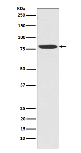
| WB, IHC, IF, ICC, IHF |
|---|---|
| Primary Accession | Q9UHD2 |
| Reactivity | Rat, Human, Mouse |
| Clonality | Monoclonal |
| Other Names | NAK; T2K; NF-kappa-B-activating kinase; TANK-binding kinase 1; Serine/threonine-protein kinase TBK1; T2K; TANK binding kinase 1; |
| Isotype | Rabbit IgG |
| Host | Rabbit |
| Calculated MW | 83642 Da |
| Dilution | WB 1:500~1:2000 IHC 1:50~1:200 ICC/IF 1:50~1:200 |
|---|---|
| Purification | Affinity-chromatography |
| Immunogen | A synthesized peptide derived from human NAK/TBK1 (N-term) |
| Description | The NF-kappa-B (NFKB) complex of proteins is inhibited by I-kappa-B (IKB) proteins, which inactivate NFKB by trapping it in the cytoplasm. Phosphorylation of serine residues on the IKB proteins by IKB kinases marks them for destruction via the ubiquitination pathway, thereby allowing activation and nuclear translocation of the NFKB complex. |
| Storage Condition and Buffer | Rabbit IgG in phosphate buffered saline , pH 7.4, 150mM NaCl, 0.02% sodium azide and 50% glycerol. Store at +4°C short term. Store at -20°C long term. Avoid freeze / thaw cycle. |
| Name | TBK1 {ECO:0000303|PubMed:10581243, ECO:0000312|HGNC:HGNC:11584} |
|---|---|
| Function | Serine/threonine kinase that plays an essential role in regulating inflammatory responses to foreign agents (PubMed:10581243, PubMed:11839743, PubMed:12692549, PubMed:12702806, PubMed:14703513, PubMed:15367631, PubMed:15485837, PubMed:18583960, PubMed:21138416, PubMed:23453971, PubMed:23453972, PubMed:23746807, PubMed:25636800, PubMed:26611359, PubMed:32404352, PubMed:34363755, PubMed:32298923). Following activation of toll-like receptors by viral or bacterial components, associates with TRAF3 and TANK and phosphorylates interferon regulatory factors (IRFs) IRF3 and IRF7 as well as DDX3X (PubMed:12692549, PubMed:12702806, PubMed:14703513, PubMed:15367631, PubMed:18583960, PubMed:25636800). This activity allows subsequent homodimerization and nuclear translocation of the IRFs leading to transcriptional activation of pro-inflammatory and antiviral genes including IFNA and IFNB (PubMed:12702806, PubMed:15367631, PubMed:25636800, PubMed:32972995). In order to establish such an antiviral state, TBK1 form several different complexes whose composition depends on the type of cell and cellular stimuli (PubMed:23453971, PubMed:23453972, PubMed:23746807). Plays a key role in IRF3 activation: acts by first phosphorylating innate adapter proteins MAVS, STING1 and TICAM1 on their pLxIS motif, leading to recruitment of IRF3, thereby licensing IRF3 for phosphorylation by TBK1 (PubMed:25636800, PubMed:30842653, PubMed:37926288). Phosphorylated IRF3 dissociates from the adapter proteins, dimerizes, and then enters the nucleus to induce expression of interferons (PubMed:25636800). Thus, several scaffolding molecules including FADD, TRADD, MAVS, AZI2, TANK or TBKBP1/SINTBAD can be recruited to the TBK1-containing- complexes (PubMed:21931631). Under particular conditions, functions as a NF-kappa-B effector by phosphorylating NF-kappa-B inhibitor alpha/NFKBIA, IKBKB or RELA to translocate NF-Kappa-B to the nucleus (PubMed:10783893, PubMed:15489227). Restricts bacterial proliferation by phosphorylating the autophagy receptor OPTN/Optineurin on 'Ser-177', thus enhancing LC3 binding affinity and antibacterial autophagy (PubMed:21617041). Phosphorylates SMCR8 component of the C9orf72-SMCR8 complex, promoting autophagosome maturation (PubMed:27103069). Phosphorylates ATG8 proteins MAP1LC3C and GABARAPL2, thereby preventing their delipidation and premature removal from nascent autophagosomes (PubMed:31709703). Seems to play a role in energy balance regulation by sustaining a state of chronic, low-grade inflammation in obesity, which leads to a negative impact on insulin sensitivity (By similarity). Attenuates retroviral budding by phosphorylating the endosomal sorting complex required for transport-I (ESCRT-I) subunit VPS37C (PubMed:21270402). Phosphorylates Borna disease virus (BDV) P protein (PubMed:16155125). Plays an essential role in the TLR3- and IFN- dependent control of herpes virus HSV-1 and HSV-2 infections in the central nervous system (PubMed:22851595). Acts both as a positive and negative regulator of the mTORC1 complex, depending on the context: activates mTORC1 in response to growth factors by catalyzing phosphorylation of MTOR, while it limits the mTORC1 complex by promoting phosphorylation of RPTOR (PubMed:29150432, PubMed:31530866). Acts as a positive regulator of the mTORC2 complex by mediating phosphorylation of MTOR, leading to increased phosphorylation and activation of AKT1 (By similarity). Phosphorylates and activates AKT1 (PubMed:21464307). Involved in the regulation of TNF-induced RIPK1- mediated cell death, probably acting via CYLD phosphorylation that in turn controls RIPK1 ubiquitination status (PubMed:34363755). Also participates in the differentiation of T follicular regulatory cells together with the receptor ICOS (PubMed:27135603). |
| Cellular Location | Cytoplasm. Note=Upon mitogen stimulation or triggering of the immune system, TBK1 is recruited to the exocyst by EXOC2. |
| Tissue Location | Ubiquitous with higher expression in testis. Expressed in the ganglion cells, nerve fiber layer and microvasculature of the retina. |
Research Areas
For Research Use Only. Not For Use In Diagnostic Procedures.
Application Protocols
Provided below are standard protocols that you may find useful for product applications.
终于等到您。ABCEPTA(百远生物)抗体产品。
点击下方“我要评价 ”按钮提交您的反馈信息,您的反馈和评价是我们最宝贵的财富之一,
我们将在1-3个工作日内处理您的反馈信息。
如有疑问,联系:0512-88856768 tech-china@abcepta.com.
¥ 1,500.00
Cat# AP90202


