


 癌症的基本特征包括细胞增殖、血管生成、迁移、凋亡逃避机制和细胞永生等。找到癌症发生过程中这些通路的关键标记物和对应的抗体用于检测至关重要。
 为您推荐一个泛素化位点预测神器——泛素化分析工具,可以为您的蛋白的泛素化位点作出预测和评分。
 细胞自噬受体图形绘图工具为你的蛋白的细胞受体结合位点作出预测和评分,识别结合到自噬通路中的蛋白是非常重要的,便于让我们理解自噬在正常生理、病理过程中的作用,如发育、细胞分化、神经退化性疾病、压力条件下、感染和癌症。
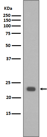
Bak Antibody
Rabbit mAb


- 产品详情
- 实验流程
Application
| WB, IHC, IF, FC, ICC, IP, IHF |
|---|---|
| Primary Accession | Q16611 |
| Reactivity | Human |
| Clonality | Monoclonal |
| Other Names | BAK1;BAK;BAK-LIKE;BCL2L7;CDN1; |
| Isotype | Rabbit IgG |
| Host | Rabbit |
| Calculated MW | 23409 Da |
| Dilution | WB 1:500~1:2000 IHC 1:50~1:200 ICC/IF 1:50~1:200 IP 1:50 FC 1:50 |
|---|---|
| Purification | Affinity-chromatography |
| Immunogen | A synthesized peptide derived from human Bak |
| Description | Bak is a proapoptotic member of the Bcl-2 family. This protein is located on the outer membrane of mitochondria and is an essential component for transduction of apoptotic signals through the mitochondrial pathway. Upon apoptotic stimulation, an upstream stimulator like truncated BID (tBID) induces conformational changes in Bak to form oligomer channels in the mitochondrial membrane for cytochrome c release. The release of cytochrome c to the cytosol activates the caspase-9 pathway and eventually leads to cell death. |
| Storage Condition and Buffer | Rabbit IgG in phosphate buffered saline , pH 7.4, 150mM NaCl, 0.02% sodium azide and 50% glycerol. Store at +4°C short term. Store at -20°C long term. Avoid freeze / thaw cycle. |
| Name | BAK1 |
|---|---|
| Synonyms | BAK, BCL2L7, CDN1 |
| Function | Plays a role in the mitochondrial apoptotic process. Upon arrival of cell death signals, promotes mitochondrial outer membrane (MOM) permeabilization by oligomerizing to form pores within the MOM. This releases apoptogenic factors into the cytosol, including cytochrome c, promoting the activation of caspase 9 which in turn processes and activates the effector caspases. |
| Cellular Location | Mitochondrion outer membrane; Single-pass membrane protein |
| Tissue Location | Expressed in a wide variety of tissues, with highest levels in the heart and skeletal muscle |
Research Areas
For Research Use Only. Not For Use In Diagnostic Procedures.
Application Protocols
Provided below are standard protocols that you may find useful for product applications.
终于等到您。ABCEPTA(百远生物)抗体产品。
点击下方“我要评价 ”按钮提交您的反馈信息,您的反馈和评价是我们最宝贵的财富之一,
我们将在1-3个工作日内处理您的反馈信息。
如有疑问,联系:0512-88856768 tech-china@abcepta.com.
¥ 1,500.00
Cat# AP90411


