


 癌症的基本特征包括细胞增殖、血管生成、迁移、凋亡逃避机制和细胞永生等。找到癌症发生过程中这些通路的关键标记物和对应的抗体用于检测至关重要。
 为您推荐一个泛素化位点预测神器——泛素化分析工具,可以为您的蛋白的泛素化位点作出预测和评分。
 细胞自噬受体图形绘图工具为你的蛋白的细胞受体结合位点作出预测和评分,识别结合到自噬通路中的蛋白是非常重要的,便于让我们理解自噬在正常生理、病理过程中的作用,如发育、细胞分化、神经退化性疾病、压力条件下、感染和癌症。
RPS3 Antibody
Rabbit mAb

- 产品详情
- 实验流程
Application
| WB, IHC, IF, ICC, IHF |
|---|---|
| Primary Accession | P23396 |
| Reactivity | Rat, Human, Mouse |
| Clonality | Monoclonal |
| Other Names | RPS3; 40S ribosomal protein S3; |
| Isotype | Rabbit IgG |
| Host | Rabbit |
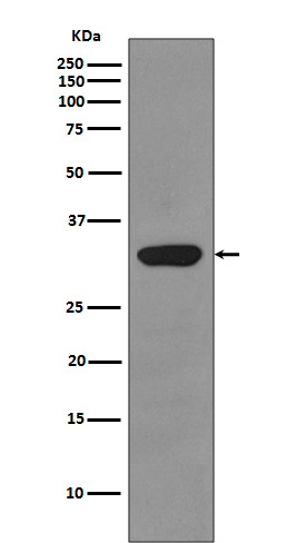
| Calculated MW | 27 KDa |
| Dilution | WB 1:500~1:2000 IHC 1:50~1:200 ICC/IF 1:50~1:200 |
|---|---|
| Purification | Affinity-chromatography |
| Immunogen | A synthesized peptide derived from human RPS3 |
| Description | Involved in translation as a component of the 40S small ribosomal subunit. Has endonuclease activity and plays a role in repair of damaged DNA. Cleaves phosphodiester bonds of DNAs containing altered bases with broad specificity and cleaves supercoiled DNA more efficiently than relaxed DNA. Displays high binding affinity for 7,8-dihydro-8-oxoguanine (8-oxoG), a common DNA lesion caused by reactive oxygen species (ROS). Has also been shown to bind with similar affinity to intact and damaged DNA. |
| Storage Condition and Buffer | Rabbit IgG in phosphate buffered saline , pH 7.4, 150mM NaCl, 0.02% sodium azide and 50% glycerol. Store at +4°C short term. Store at -20°C long term. Avoid freeze / thaw cycle. |
Research Areas
For Research Use Only. Not For Use In Diagnostic Procedures.
Application Protocols
Provided below are standard protocols that you may find useful for product applications.
终于等到您。ABCEPTA(百远生物)抗体产品。
点击下方“我要评价 ”按钮提交您的反馈信息,您的反馈和评价是我们最宝贵的财富之一,
我们将在1-3个工作日内处理您的反馈信息。
如有疑问,联系:0512-88856768 tech-china@abcepta.com.
¥ 1,500.00
Cat# AP90526


