


 癌症的基本特征包括细胞增殖、血管生成、迁移、凋亡逃避机制和细胞永生等。找到癌症发生过程中这些通路的关键标记物和对应的抗体用于检测至关重要。
 为您推荐一个泛素化位点预测神器——泛素化分析工具,可以为您的蛋白的泛素化位点作出预测和评分。
 细胞自噬受体图形绘图工具为你的蛋白的细胞受体结合位点作出预测和评分,识别结合到自噬通路中的蛋白是非常重要的,便于让我们理解自噬在正常生理、病理过程中的作用,如发育、细胞分化、神经退化性疾病、压力条件下、感染和癌症。
NALP3 Antibody
Rabbit mAb


- 产品详情
- 实验流程
Application
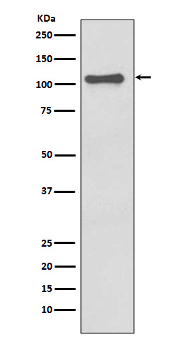
| WB, IHC, IF, ICC, IHF |
|---|---|
| Primary Accession | Q96P20 |
| Reactivity | Rat, Human, Mouse |
| Clonality | Monoclonal |
| Other Names | FCU; MWS; FCAS; Cias1; Mmig1; NLRP3; Pypaf1; AII/AVP; AGTAVPRL; Cryopyrin; |
| Isotype | Rabbit IgG |
| Host | Rabbit |
| Calculated MW | 118173 Da |
| Dilution | WB 1:500~1:2000 IHC 1:50~1:200 ICC/IF 1:50~1:200 |
|---|---|
| Purification | Affinity-chromatography |
| Immunogen | A synthesized peptide derived from human NALP3 |
| Description | As the sensor component of the NLRP3 inflammasome, plays a crucial role in innate immunity and inflammation. |
| Storage Condition and Buffer | Rabbit IgG in phosphate buffered saline , pH 7.4, 150mM NaCl, 0.02% sodium azide and 50% glycerol. Store at +4°C short term. Store at -20°C long term. Avoid freeze / thaw cycle. |
| Name | NLRP3 {ECO:0000303|PubMed:17907925, ECO:0000312|HGNC:HGNC:16400} |
|---|---|
| Function | Sensor component of the NLRP3 inflammasome, which mediates inflammasome activation in response to defects in membrane integrity, leading to secretion of inflammatory cytokines IL1B and IL18 and pyroptosis (PubMed:16407889, PubMed:18403674, PubMed:18604214, PubMed:23582325, PubMed:25686105, PubMed:27929086, PubMed:28656979, PubMed:28847925, PubMed:30487600, PubMed:30612879, PubMed:31086327, PubMed:31086329, PubMed:31189953, PubMed:33231615, PubMed:34133077, PubMed:34341353, PubMed:34512673, PubMed:36442502, PubMed:40450990). In response to pathogens and other damage-associated signals that affect the integrity of membranes, initiates the formation of the inflammasome polymeric complex composed of NLRP3, CASP1 and PYCARD/ASC (PubMed:16407889, PubMed:18403674, PubMed:27432880, PubMed:28847925, PubMed:31189953, PubMed:33231615, PubMed:34133077, PubMed:34341353, PubMed:36142182, PubMed:36442502). Recruitment of pro-caspase-1 (proCASP1) to the NLRP3 inflammasome promotes caspase-1 (CASP1) activation, which subsequently cleaves and activates inflammatory cytokines IL1B and IL18 and gasdermin-D (GSDMD), promoting cytokine secretion and pyroptosis (PubMed:23582325, PubMed:28847925, PubMed:31189953, PubMed:33231615, PubMed:34133077, PubMed:34341353). Activation of NLRP3 inflammasome is also required for HMGB1 secretion; stimulating inflammatory responses (PubMed:22801494). Involved in the homeostatic wound healing response to tissue injury, a multistep cascade that guides neutrophil migration to necrotic sites while avoiding collateral damage of healthy tissues. ATP released from necrotic cells triggers activation of NLRP3 inflammasome through P2RX7 signaling leading to neutrophil adhesion to the vascular endothelium close to the injury site (By similarity). Under resting conditions, ADP-bound NLRP3 is autoinhibited (PubMed:35114687). NLRP3 activation stimuli include extracellular ATP, nigericin, reactive oxygen species, crystals of monosodium urate or cholesterol, amyloid-beta fibers, environmental or industrial particles and nanoparticles, such as asbestos, silica, aluminum salts, cytosolic dsRNA, etc (PubMed:16407889, PubMed:18403674, PubMed:18604214, PubMed:19414800, PubMed:23871209). Almost all stimuli trigger intracellular K(+) efflux (By similarity). These stimuli lead to membrane perturbation and activation of NLRP3 (By similarity). Upon activation, NLRP3 is transported to microtubule organizing center (MTOC), where it is unlocked by NEK7, leading to its relocalization to dispersed trans- Golgi network (dTGN) vesicle membranes and formation of an active inflammasome complex (PubMed:36442502, PubMed:39173637). Associates with dTGN vesicle membranes by binding to phosphatidylinositol 4- phosphate (PtdIns4P) (PubMed:30487600, PubMed:34554188). Shows ATPase activity (PubMed:17483456). |
| Cellular Location | Cytoplasm, cytosol. Inflammasome. Cytoplasm, cytoskeleton, microtubule organizing center. Golgi apparatus membrane. Endoplasmic reticulum {ECO:0000250|UniProtKB:Q8R4B8}. Mitochondrion. Secreted. Nucleus {ECO:0000250|UniProtKB:Q8R4B8} Note=In macrophages, under resting conditions, mainly located in the cytosol and on membranes of various organelles, such as endoplasmic reticulum, mitochondria and Golgi: forms an inactive double-ring cage that is primarily localized on membranes (By similarity). Upon activation, NLRP3 is transported to microtubule organizing center (MTOC), where it is unlocked by NEK7, leading to its relocalization to dispersed trans-Golgi network (dTGN) vesicle membranes for the formation of an active inflammasome complex (PubMed:39173637) Recruited to dTGN vesicle membranes by binding to phosphatidylinositol 4-phosphate (PtdIns4P) (PubMed:30487600). After the induction of pyroptosis, inflammasome specks are released into the extracellular space where they can further promote IL1B processing and where they can be engulfed by macrophages (PubMed:24952504). Phagocytosis induces lysosomal damage and inflammasome activation in the recipient cells (PubMed:24952504). In the Th2 subset of CD4(+) helper T cells, mainly located in the nucleus (By similarity). Nuclear localization depends upon KPNA2 (By similarity). In the Th1 subset of CD4(+) helper T cells, mainly cytoplasmic (By similarity). {ECO:0000250|UniProtKB:Q8R4B8, ECO:0000269|PubMed:24952504, ECO:0000269|PubMed:30487600, ECO:0000269|PubMed:39173637} |
| Tissue Location | Predominantly expressed in macrophages (PubMed:33231615, PubMed:34133077). Also expressed in dendritic cells, B- and T-cells (at protein level) (PubMed:11786556, PubMed:17164409) Expressed in LPS-treated granulocytes, but not in resting cells (at protein level) (PubMed:17164409). Expression in monocytes is very weak (at protein level) (PubMed:17164409). Expressed in stratified non- keratinizing squamous epithelium, including oral, esophageal and ectocervical mucosa and in the Hassall's corpuscles in the thymus Also, detected in the stratified epithelium covering the bladder and ureter (transitional mucosa) (at protein level) (PubMed:17164409) Expressed in lung epithelial cells (at protein level) (PubMed:23229815). Expressed in chondrocytes (PubMed:12032915) Expressed at low levels in resting osteoblasts (PubMed:17907925) |
Research Areas
For Research Use Only. Not For Use In Diagnostic Procedures.
Application Protocols
Provided below are standard protocols that you may find useful for product applications.
终于等到您。ABCEPTA(百远生物)抗体产品。
点击下方“我要评价 ”按钮提交您的反馈信息,您的反馈和评价是我们最宝贵的财富之一,
我们将在1-3个工作日内处理您的反馈信息。
如有疑问,联系:0512-88856768 tech-china@abcepta.com.
¥ 1,500.00
Cat# AP90654


